
MTSS oder LIMP? Warum wir das „Schienbeinkantensyndrom“ neu denken müssen
Ein vertrautes Beschwerdebild – mit mehr Fragezeichen als gedacht
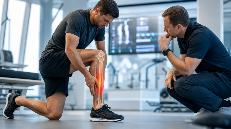
Laufen ist populär wie selten zuvor. Gleichzeitig gehört der belastungsabhängige Schmerz an der medialen Tibiakante zu den hartnäckigsten Überlastungsproblemen des Unterschenkels. Das sogenannte „Schienbeinkantensyndrom“ oder Medial Tibial Stress Syndrome (MTSS) kann Läufer über Wochen aus dem Training nehmen. Die Rückkehr zum Sport verlangt deshalb mehr als die einfache Empfehlung, zunächst zu pausieren.
Doch schon die Bezeichnung wirft Fragen auf: Beschreibt MTSS tatsächlich die zugrunde liegende Pathophysiologie? Oder wäre Load Induced Medial-Leg Pain (LIMP) eine präzisere Bezeichnung, die Schmerzort und Belastungsbezug beschreibt, ohne vorschnell eine bestimmte Gewebestruktur als Ursache festzulegen?
Häufig – aber ursächlich weiterhin unklar
Die epidemiologischen Daten schwanken erheblich. Je nach Population wurden Prävalenzen zwischen 5,4 und 69,5 Prozent sowie Inzidenzen von etwa 16 bis 56 Prozent berichtet. Besonders häufig tritt das Beschwerdebild unter Militärrekrut und bestimmten Läufergruppen auf.
Trotz der hohen Häufigkeit ist die Ursache nicht eindeutig geklärt. Frühere Theorien stellten Periost, Faszien und Sehnen in den Mittelpunkt. Neuere Befunde sprechen teilweise für eine Beteiligung des Knochens und eine Einordnung als Knochenstressverletzung. Allerdings sind auch diese Befunde nicht eindeutig und können teilweise physiologische Anpassungsreaktionen auf erhöhte Belastung darstellen.
Damit wird ein zentrales Problem sichtbar: Wir wissen, dass Belastung eine entscheidende Rolle spielt – aber nicht abschließend, welches Gewebe den Schmerz verursacht.
Risikofaktoren: Hinweise statt einfache Ursachen
Untersucht wurden zahlreiche mögliche Risikofaktoren. Dazu zählen unter anderem ein höherer BMI, ein größerer Navicular Drop sowie bestimmte Bewegungsumfänge von Sprunggelenk und Hüfte. Auch Lauf- und Bewegungsmuster, Fußgewölbe, Druckverteilung, Soleusaktivität sowie Hüft- und Rumpfkontrolle wurden mit MTSS in Verbindung gebracht.
Die Interpretation dieser Befunde verlangt jedoch Vorsicht. Ein größerer Navicular Drop kann beispielsweise mit MTSS assoziiert sein, der klassische Test besitzt aber Einschränkungen hinsichtlich Reliabilität und bildet dynamische Bewegungen beim Laufen nur begrenzt ab. Ähnliches gilt für biomechanische Auffälligkeiten: Sie können Ursache, Folge der Schmerzen oder lediglich Begleiterscheinung sein.
Der gemeinsame Nenner bleibt daher ein Missverhältnis zwischen mechanischer Belastung und Gewebetoleranz.
Warum LIMP einen Perspektivwechsel ermöglichen könnte
Anderson et al. schlagen 2025 vor, MTSS durch Load Induced Medial-Leg Pain (LIMP) zu ersetzen. Der Begriff benennt den auslösenden Faktor und den Schmerzort, ohne eine ungeklärte Pathologie vorwegzunehmen. Das könnte die klinische Kommunikation verändern: weg von der vermeintlich eindeutigen Gewebediagnose, hin zu einer belastungsorientierten Betrachtung.
Diagnostik: Erst differenzieren, dann behandeln
Typisch sind diffuse, belastungsinduzierte Schmerzen entlang der posteromedialen Tibiakante über mehr als fünf Zentimeter, die durch Palpation reproduzierbar sind. Wichtig ist die Abgrenzung zu anderen Ursachen wie Stressfraktur oder chronischem belastungsinduziertem Kompartmentsyndrom. Je nach klinischem Verdacht kann Bildgebung erforderlich sein.
Therapie: Belastung statt Stillstand
Die Evidenz zur Behandlung ist begrenzt. Als zentrale konservative Strategie gilt eine graduelle Belastungssteuerung, begleitet von Patientenedukation und realistischem Erwartungsmanagement. Die Rückkehr zum Laufen kann deutlich länger dauern als erwartet; bei länger bestehenden Beschwerden werden Heilungszeiträume von mehreren Monaten beschrieben.
Auch Laufprogramme, Kräftigung, Dehnung, Kompressionssocken, Einlagen oder Stoßwellentherapie wurden untersucht. Die Studienlage ist jedoch heterogen und teilweise methodisch schwach. Operative Verfahren sollten aufgrund der geringen Evidenz und des unklaren Pathomechanismus nicht als erste Option betrachtet werden.
Der entscheidende Gedanke
Die wichtigste Konsequenz lautet deshalb nicht einfach „MTSS behandeln“, sondern Belastung und Gewebetoleranz wieder zusammenzubringen. LIMP könnte hierfür eine Sprache liefern, die klinische Unsicherheit ausdrücklich zulässt und dennoch konkrete Therapieentscheidungen ermöglicht.
Doch genau hier beginnt die eigentlich spannende Frage: Wenn wir den Begriff verändern, verändert sich dann auch unser klinisches Denken – und damit möglicherweise unsere gesamte Herangehensweise an Diagnostik, Belastungssteuerung und Rückkehr zum Laufen?
Die vollständige Academy-Version zeigt, wie sich aus dieser Perspektive konkrete klinische Entscheidungen ableiten lassen – und wo die aktuelle Evidenz dabei klare Grenzen setzt.
Dein DK Sports & Physio Team aus der Karlsruher Oststadt
Den ausführlichen Artikel findest du in unserer DK Academy.
Wir geben Physiotherapeuten, Trainern und allen Wissbegierigen einen sachlichen Einblick in die Physiotherapie und helfen so die Rehabilitation und das Training nach Verletzungen oder Beschwerden effizienter zu gestalten.
Sichere dir vollen Zugriff auf unsere Rehab Live Sessions, exklusive Review- und Blogartikel, Simple Tipps und Infografiken.

Du benötigst Physiotherapie im Raum Karlsruhe? Dann sind wir gerne für dich da und unterstützen dich!
Dein DK Sports & Physio Team aus der Karlsruher Oststadt
Unsere weiteren Blog-Artikel

Training als Medikament bei Multipler Sklerose?

Vegane Ernährung im Leistungssport – zwischen Planung, Evidenz und Performance
MTSS oder LIMP? Warum wir das „Schienbeinkantensyndrom“ neu denken müssen

Glutenunverträglichkeit – alles nur Kopfsache?

Airex-Kissen & Bosu-Bälle – Spielzeug oder kann das weg?
Warum wir über die Reruptur des Kreuzbandes sprechen müssen

Arroganz in der Physiotherapie? Zwischen Wissen und Gewissheit
Quellenangaben:
[1] D. Van Poppel et al., “Risk factors for overuse injuries in short- and long-distance running: A systematic review,” Journal of Sport and Health Science, vol. 10, no. 1, pp. 14–28, Jan. 2021, doi: 10.1016/j.jshs.2020.06.006.
[2] A. Vitti, P. T. Nikolaidis, E. Villiger, V. Onywera, and B. Knechtle, “The ‘New York City Marathon’: participation and performance trends of 1.2M runners during half-century,” Research in Sports Medicine, vol. 28, no. 1, pp. 121–137, Jan. 2020, doi: 10.1080/15438627.2019.1586705.
[3] S. W. Bredeweg, S. Zijlstra, B. Bessem, and I. Buist, “The effectiveness of a preconditioning programme on preventing running-related injuries in novice runners: a randomised controlled trial,” Br J Sports Med, vol. 46, no. 12, pp. 865–870, Sep. 2012, doi: 10.1136/bjsports-2012-091397.
[4] N. Kakouris, N. Yener, and D. T. P. Fong, “A systematic review of running-related musculoskeletal injuries in runners,” Journal of Sport and Health Science, vol. 10, no. 5, pp. 513–522, Sep. 2021, doi: 10.1016/j.jshs.2021.04.001.
[5] C. Menéndez, L. Batalla, A. Prieto, M. Á. Rodríguez, I. Crespo, and H. Olmedillas, “Medial Tibial Stress Syndrome in Novice and Recreational Runners: A Systematic Review,” IJERPH, vol. 17, no. 20, p. 7457, Oct. 2020, doi: 10.3390/ijerph17207457.
[6] B. Mulvad, R. O. Nielsen, M. Lind, and D. Ramskov, “Diagnoses and time to recovery among injured recreational runners in the RUN CLEVER trial,” PLoS ONE, vol. 13, no. 10, p. e0204742, Oct. 2018, doi: 10.1371/journal.pone.0204742.
[7] M. Franklyn, “Aetiology and mechanisms of injury in medial tibial stress syndrome: Current and future developments,” WJO, vol. 6, no. 8, p. 577, 2015, doi: 10.5312/wjo.v6.i8.577.
[8] M. A. Saad, J. M. Jamal, A. T. Aldhafiri, and S. A. Alkandari, “Medial Tibial Stress Syndrome: A Scoping Review of Epidemiology, Biomechanics, and Risk Factors,” Cureus, Mar. 2025, doi: 10.7759/cureus.81463.
[9] Prina. Y. Patel and N. Patil, “PREVALENCE OF SHIN SPLINT IN RECREATIONAL MARATHON RUNNER,” International Journal of Physiotherapy, vol. 7, no. 1, Feb. 2020, doi: 10.15621/ijphy/2020/v7i1/193672.
[10] A. Chandran, S. N. Morris, P. R. Roby, A. J. Boltz, H. J. Robison, and C. L. Collins, “Epidemiology of Injuries in National Collegiate Athletic Association Women’s Track and Field: 2014–2015 Through 2018–2019,” Journal of Athletic Training, vol. 56, no. 7, pp. 780–787, Jul. 2021, doi: 10.4085/1062-6050-493-20.
[11] K. Knobloch, U. Yoon, and P. M. Vogt, “Acute and Overuse Injuries Correlated to Hours of Training in Master Running Athletes,” Foot Ankle Int., vol. 29, no. 7, pp. 671–676, Jul. 2008, doi: 10.3113/FAI.2008.0671.
[12] M. Alessa et al., “Highlights of Medial Tibial Stress Syndrome in Military Recruits: A Narrative Review,” Cureus, Dec. 2024, doi: 10.7759/cureus.75376.
[13] M. H. Moen, J. L. Tol, A. Weir, M. Steunebrink, and T. C. De Winter, “Medial Tibial Stress Syndrome: A Critical Review,” Sports Medicine, vol. 39, no. 7, pp. 523–546, Jul. 2009, doi: 10.2165/00007256-200939070-00002.
[14] T. Okunuki et al., “Relationship between medial tibial stress syndrome and the adipose tissue along the posteromedial tibial border and the crural chiasma,” The Physician and Sportsmedicine, vol. 53, no. 4, pp. 299–308, Jul. 2025, doi: 10.1080/00913847.2025.2462979.
[15] M. Winters, P. Bon, S. Bijvoet, E. W. P. Bakker, and M. H. Moen, “Are ultrasonographic findings like periosteal and tendinous edema associated with medial tibial stress syndrome? A case-control study,” Journal of Science and Medicine in Sport, vol. 20, no. 2, pp. 128–133, Feb. 2017, doi: 10.1016/j.jsams.2016.07.001.
[16] T. Hoenig et al., “International Delphi consensus on bone stress injuries in athletes,” Br J Sports Med, vol. 59, no. 2, pp. 78–90, Jan. 2025, doi: 10.1136/bjsports-2024-108616.
[17] M. Gaeta et al., “High-Resolution CT Grading of Tibial Stress Reactions in Distance Runners,” American Journal of Roentgenology, vol. 187, no. 3, pp. 789–793, Sep. 2006, doi: 10.2214/AJR.05.0303.
[18] M. Winters, D. B. Burr, H. Van Der Hoeven, K. W. Condon, J. Bellemans, and M. H. Moen, “Microcrack-associated bone remodeling is rarely observed in biopsies from athletes with medial tibial stress syndrome,” J Bone Miner Metab, vol. 37, no. 3, pp. 496–502, May 2019, doi: 10.1007/s00774-018-0945-9.
[19] L. A. Weidauer, M. M. Eilers, T. L. Binkley, M. D. Vukovich, and B. L. Specker, “Effect of different collegiate sports on cortical bone in the tibia,” J Musculoskelet Neuronal Interact, vol. 12, no. 2, pp. 68–73, Jun. 2012.
[20] K. L. Hamstra-Wright, K. C. H. Bliven, and C. Bay, “Risk factors for medial tibial stress syndrome in physically active individuals such as runners and military personnel: a systematic review and meta-analysis,” Br J Sports Med, vol. 49, no. 6, pp. 362–369, Mar. 2015, doi: 10.1136/bjsports-2014-093462.
[21] D. M. Brody, “Techniques in the evaluation and treatment of the injured runner,” Orthop Clin North Am, vol. 13, no. 3, pp. 541–558, Jul. 1982.
[22] M. F. Alam, S. Zaki, S. Sharma, M. Ghareeb, and S. Nuhmani, “Intra-rater reliability and validity of navicular drop test for the assessment of medial longitudinal arch in patients having pronated feet with chronic low back pain,” Clinical Epidemiology and Global Health, vol. 32, p. 101939, Mar. 2025, doi: 10.1016/j.cegh.2025.101939.
[23] A. Vinicombe, A. Raspovic, and H. B. Menz, “Reliability of Navicular Displacement Measurement as a Clinical Indicator of Foot Posture,” Journal of the American Podiatric Medical Association, vol. 91, no. 5, pp. 262–268, May 2001, doi: 10.7547/87507315-91-5-262.
[24] M. S. Rathleff, R. G. Nielsen, and U. G. Kersting, “Navicula Drop Test Ad Modum Brody: Does It Show How the Foot Moves Under Dynamic Conditions?,” J Am Podiatr Med Assoc, vol. 102, no. 1, pp. 34–38, Jan. 2012, doi: 10.7547/1020034.
[25] T. J. Hubbard, E. M. Carpenter, and M. L. Cordova, “Contributing Factors to Medial Tibial Stress Syndrome: A Prospective Investigation,” Medicine & Science in Sports & Exercise, vol. 41, no. 3, pp. 490–496, Mar. 2009, doi: 10.1249/MSS.0b013e31818b98e6.
[26] A. Naderi, M. H. Moen, and H. Degens, “Is high soleus muscle activity during the stance phase of the running cycle a potential risk factor for the development of medial tibial stress syndrome? A prospective study,” Journal of Sports Sciences, vol. 38, no. 20, pp. 2350–2358, Oct. 2020, doi: 10.1080/02640414.2020.1785186.
[27] B. Noh, A. Masunari, K. Akiyama, M. Fukano, T. Fukubayashi, and S. Miyakawa, “Structural deformation of longitudinal arches during running in soccer players with medial tibial stress syndrome,” European Journal of Sport Science, vol. 15, no. 2, pp. 173–181, Mar. 2015, doi: 10.1080/17461391.2014.932848.
[28] J. Becker, M. Nakajima, and W. F. W. Wu, “Factors Contributing to Medial Tibial Stress Syndrome in Runners: A Prospective Study,” Medicine & Science in Sports & Exercise, vol. 50, no. 10, pp. 2092–2100, Oct. 2018, doi: 10.1249/MSS.0000000000001674.
[29] V. Sobhani, A. Shakibaee, A. Khatibi Aghda, M. K. Emami Meybodi, A. Delavari, and D. Jahandideh, “Studying the Relation Between Medial Tibial Stress Syndrome and Anatomic and Anthropometric Characteristics of Military Male Personnel,” Asian J Sports Med, vol. 6, no. 1, Mar. 2015, doi: 10.5812/asjsm.23811.
[30] M. S. Rathleff, L. A. Kelly, F. B. Christensen, O. H. Simonsen, S. Kaalund, and U. Laessoe, “Dynamic Midfoot Kinematics in Subjects with Medial Tibial Stress Syndrome,” J Am Podiatr Med Assoc, vol. 102, no. 3, pp. 205–212, May 2012, doi: 10.7547/1020205.
[31] M. H. Moen et al., “Risk factors and prognostic indicators for medial tibial stress syndrome,” Scandinavian Med Sci Sports, vol. 22, no. 1, pp. 34–39, Feb. 2012, doi: 10.1111/j.1600-0838.2010.01144.x.
[32] J. L. Tweed, J. A. Campbell, and S. J. Avil, “Biomechanical Risk Factors in the Development of Medial Tibial Stress Syndrome in Distance Runners,” JAPMA, vol. 98, no. 6, pp. 436–444, Nov. 2008, doi: 10.7547/0980436.
[33] B. Yates and S. White, “The Incidence and Risk Factors in the Development of Medial Tibial Stress Syndrome among Naval Recruits,” Am J Sports Med, vol. 32, no. 3, pp. 772–780, Apr. 2004, doi: 10.1177/0095399703258776.
[34] C. Bramah, S. J. Preece, N. Gill, and L. Herrington, “Is There a Pathological Gait Associated With Common Soft Tissue Running Injuries?,” Am J Sports Med, vol. 46, no. 12, pp. 3023–3031, Oct. 2018, doi: 10.1177/0363546518793657.
[35] L. M. Anderson, S. J. Warden, R. W. Willy, D. R. Bonanno, and H. B. Menz, “Medial Tibial Stress Syndrome Needs a New Name—Make No Bones About It,” Journal of Orthopaedic & Sports Physical Therapy, vol. 55, no. 10, pp. 621–622, Oct. 2025, doi: 10.2519/jospt.2025.13411.
[36] M. Winters, E. W. P. Bakker, M. H. Moen, C. C. Barten, R. Teeuwen, and A. Weir, “Medial tibial stress syndrome can be diagnosed reliably using history and physical examination,” Br J Sports Med, vol. 52, no. 19, pp. 1267–1272, Oct. 2018, doi: 10.1136/bjsports-2016-097037.
[37] H. Lohrer, N. Malliaropoulos, V. Korakakis, and N. Padhiar, “Exercise-induced leg pain in athletes: diagnostic, assessment, and management strategies,” The Physician and Sportsmedicine, vol. 47, no. 1, pp. 47–59, Jan. 2019, doi: 10.1080/00913847.2018.1537861.
[38] M. Winters, “The diagnosis and management of medial tibial stress syndrome: An evidence update,” Unfallchirurg, vol. 123, no. S1, pp. 15–19, Jan. 2020, doi: 10.1007/s00113-019-0667-z.
[39] M. H. Moen et al., “The treatment of medial tibial stress syndrome in athletes; a randomized clinical trial,” BMC Sports Sci Med Rehabil, vol. 4, no. 1, p. 12, Dec. 2012, doi: 10.1186/1758-2555-4-12.
[40] M. H. Moen et al., “Shockwave treatment for medial tibial stress syndrome in athletes; a prospective controlled study,” Br J Sports Med, vol. 46, no. 4, pp. 253–257, Mar. 2012, doi: 10.1136/bjsm.2010.081992.
[41] T. B. T. Marques, R. P. S. Rangel, L. V. Martins, and A. P. D. C. Vidal, “Preventive interventions for medial tibial stress syndrome: Systematic review and meta-analysis,” Gait & Posture, vol. 122, pp. 92–98, Oct. 2025, doi: 10.1016/j.gaitpost.2025.07.312.