
Post-COVID in der Physiotherapie: Sicher behandeln trotz Unsicherheit
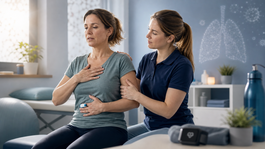
Post-COVID bleibt eine Herausforderung – auch Jahre nach der Pandemie
Für viele Menschen ist COVID-19 längst Vergangenheit. Doch für Millionen Betroffene weltweit sind die gesundheitlichen Folgen bis heute Realität. Das Post-COVID-Syndrom (PCS) kann Monate nach einer Infektion bestehen bleiben und äußert sich unter anderem durch Fatigue, Belastungsintoleranz, Atembeschwerden oder kognitive Einschränkungen. Da das Krankheitsbild noch jung ist und die Evidenz stetig wächst, stehen Physiotherapeut:innen häufig vor der Herausforderung, Patient:innen sicher zu behandeln, obwohl es bislang keine einheitlichen Standards gibt.
Dieser Beitrag gibt einen Überblick über die wichtigsten Erkenntnisse zu Definition, Risikofaktoren, Symptomen und den aktuellen Empfehlungen für eine sichere physiotherapeutische Versorgung.
Was genau ist das Post-COVID-Syndrom?
Bereits bei der Begriffsdefinition beginnt die Unsicherheit. International existieren unterschiedliche Bezeichnungen und Zeiträume für anhaltende Beschwerden nach einer SARS-CoV-2-Infektion. In Deutschland wird von einem Post-COVID-Syndrom gesprochen, wenn Symptome länger als zwölf Wochen bestehen und sich nicht anderweitig erklären lassen.
Diese fehlende Einheitlichkeit erschwert sowohl die Forschung als auch die Interpretation wissenschaftlicher Studien – ein wichtiger Aspekt für alle, die evidenzbasiert arbeiten möchten.
Wer ist betroffen?
Die Häufigkeit von PCS variiert je nach Studie erheblich. Unterschiede in Definitionen, Studienpopulationen und Impfstatus führen zu sehr unterschiedlichen Prävalenzangaben. Besonders häufig betroffen sind Frauen mittleren Alters, Menschen mit Vorerkrankungen, höherem BMI oder schwerem Akutverlauf. Dennoch kann PCS grundsätzlich nach jeder COVID-19-Infektion auftreten – auch nach milden oder sogar symptomfreien Verläufen.
Warum entstehen die Beschwerden?
Die genauen Ursachen sind bislang nicht vollständig geklärt. Vermutet wird ein Zusammenspiel verschiedener biologischer Mechanismen: anhaltende Immunaktivierung, Störungen der Energieproduktion in den Mitochondrien, Gefäßveränderungen sowie Fehlregulationen des autonomen Nervensystems. Diese Prozesse können unterschiedliche Organsysteme gleichzeitig betreffen und erklären die große Vielfalt der Symptome.
Die wichtigsten Symptome für die Physiotherapie
Im Mittelpunkt stehen häufig eine ausgeprägte Fatigue und eine eingeschränkte Belastbarkeit. Besonders relevant ist die sogenannte Post-Exertionelle Malaise (PEM) – eine verzögerte Symptomverschlechterung nach körperlicher, geistiger oder emotionaler Belastung. Hinzu kommen Atembeschwerden, Konzentrationsstörungen („Brain Fog“), Schmerzen, Schlafstörungen und autonome Dysfunktionen wie POTS.
Gerade PEM verändert den therapeutischen Umgang grundlegend: Belastung muss individuell gesteuert werden, da klassische Trainingsprinzipien bei diesen Patient:innen kontraproduktiv sein können.
Sicherheit steht an erster Stelle
Da es bislang keine eindeutigen Biomarker oder standardisierten Diagnoseverfahren gibt, bildet eine strukturierte Anamnese die Grundlage jeder Behandlung. Vor Beginn einer aktiven Therapie sollten mögliche Warnzeichen wie PEM, kardiologische Beschwerden, belastungsabhängige Sauerstoffabfälle oder Störungen des autonomen Nervensystems erkannt werden.
Nur wenn diese Faktoren berücksichtigt werden, lässt sich das Risiko einer Symptomverschlechterung minimieren.
Therapie bedeutet individuelle Belastungssteuerung
Ein universelles Therapieprotokoll existiert nicht. Stattdessen orientiert sich die Behandlung konsequent an Symptomen, Belastbarkeit und persönlichen Zielen der Betroffenen. Besonders wichtig sind eine vorsichtige Dosierung körperlicher Aktivität, ausreichende Erholungsphasen sowie Strategien des Energie- und Aktivitätsmanagements wie Pacing. Fortschritt zeigt sich häufig nicht durch Leistungssteigerung, sondern zunächst durch Stabilität und eine bessere Kontrolle des Alltags.
Die größte Herausforderung beginnt erst in der Praxis
Die wissenschaftliche Evidenz entwickelt sich kontinuierlich weiter – und mit ihr die Empfehlungen für Diagnostik und Therapie. Doch welche Screenings sollten Physiotherapeut:innen unbedingt durchführen? Wie erkennt man PEM zuverlässig? Wann ist Training sinnvoll, wann sogar kontraindiziert? Und wie lassen sich Belastung, Herzfrequenz und Progression sicher steuern, ohne Rückfälle zu provozieren?
Die vollständige Version dieses Beitrags zeigt Schritt für Schritt, wie sich aktuelle Evidenz in die physiotherapeutische Praxis übertragen lässt – inklusive konkreter Screeningverfahren, Therapiealgorithmen, Pacing-Strategien und Handlungsempfehlungen für unterschiedliche Symptomkonstellationen.
Dein DK Sports & Physio Team aus der Karlsruher Oststadt
Den ausführlichen Artikel findest du in unserer DK Academy.
Wir geben Physiotherapeuten, Trainern und allen Wissbegierigen einen sachlichen Einblick in die Physiotherapie und helfen so die Rehabilitation und das Training nach Verletzungen oder Beschwerden effizienter zu gestalten.
Sichere dir vollen Zugriff auf unsere Rehab Live Sessions, exklusive Review- und Blogartikel, Simple Tipps und Infografiken.

Du benötigst Physiotherapie im Raum Karlsruhe? Dann sind wir gerne für dich da und unterstützen dich!
Dein DK Sports & Physio Team aus der Karlsruher Oststadt
Unsere weiteren Blog-Artikel
Hallux Valgus – Nur ein Schönheitsmakel?
Krafttraining ist Medizin!

Post-COVID in der Physiotherapie: Sicher behandeln trotz Unsicherheit
Sarkopenie: Wie wir den Muskelschwund erkennen und stoppen
Die Königin aller Übungen – Die Kniebeuge
Schlaf Kindlein, Schlaf, warum guter Schlaf so wichtig ist!
Rotator cuff related shoulder pain – was steckt dahinter?
Quellenangaben:
[1] H. E. Davis, L. McCorkell, J. M. Vogel, und E. J. Topol, „Long COVID: major findings, mechanisms and recommendations“, Nat. Rev. Microbiol., Bd. 21, Nr. 3, S. 133–146, 2023, doi: 10.1038/s41579-022-00846-2.
[2] A. V. Ballering, S. K. R. van Zon, T. C. olde Hartman, und J. G. M. Rosmalen, „Persistence of somatic symptoms after COVID-19 in the Netherlands: an observational cohort study“, Lancet Lond. Engl., Bd. 400, Nr. 10350, S. 452–461, 2022, doi: 10.1016/S0140-6736(22)01214-4.
[3] J. Gutzeit u. a., „Definitions and symptoms of the post-COVID syndrome: an updated systematic umbrella review“, Eur. Arch. Psychiatry Clin. Neurosci., Bd. 275, Nr. 1, S. 129–140, 2025, doi: 10.1007/s00406-024-01868-y.
[4] World Health Organization (WHO), „Living guidance for clinical management of COVID-19“, World Health Organization (WHO), Nov. 2021. [Online]. Verfügbar unter: https://iris.who.int/server/api/core/bitstreams/670e3b89-b4ae-429b-abee-7b0d3d54af49/content
[5] National Institute for Health and Care Excellence (NICE), „COVID-19 rapid guideline: managing the long-term effects of COVID-19“, Jan. 2025. [Online]. Verfügbar unter: www.nice.org.uk/guidance/ng188
[6] CDC, „CDC Science and the Public Health Approach to Long COVID“, COVID-19. Zugegriffen: 19. Januar 2026. [Online]. Verfügbar unter: https://www.cdc.gov/covid/php/long-covid/index.html
[7] „Long COVID | NHLBI, NIH“. Zugegriffen: 19. Januar 2026. [Online]. Verfügbar unter: https://www.nhlbi.nih.gov/covid/long-covid
[8] A. R. Koczulla u. a., „S1-Leitlinie Long-/Post-COVID“, Pneumologie, Bd. 76, Nr. 12, S. 855–907, Dez. 2022, doi: 10.1055/a-1946-3230.
[9] T. L. Kelley, Interpretation of educational measurements. in Interpretation of educational measurements. Oxford, England: World Book Co., 1927, S. 353.
[10] H. Ledford, „How common is long COVID? Why studies give different answers“, Nature, Bd. 606, Nr. 7916, S. 852–853, Juni 2022, doi: 10.1038/d41586-022-01702-2.
[11] T. Greenhalgh, M. Sivan, A. Perlowski, und J. Ž. Nikolich, „Long COVID: a clinical update“, The Lancet, Bd. 404, Nr. 10453, S. 707–724, Aug. 2024, doi: 10.1016/S0140-6736(24)01136-X.
[12] B. Bowe, Y. Xie, und Z. Al-Aly, „Postacute sequelae of COVID-19 at 2 years“, Nat. Med., Bd. 29, Nr. 9, S. 2347–2357, 2023, doi: 10.1038/s41591-023-02521-2.
[13] A. Pavli, M. Theodoridou, und H. C. Maltezou, „Post-COVID Syndrome: Incidence, Clinical Spectrum, and Challenges for Primary Healthcare Professionals“, Arch. Med. Res., Bd. 52, Nr. 6, S. 575–581, Aug. 2021, doi: 10.1016/j.arcmed.2021.03.010.
[14] Office for National Statistics (ONS), „Prevalence of ongoing symptoms following coronavirus (COVID-19) infection in the UK – Office for National Statistics“. Zugegriffen: 23. Oktober 2025. [Online]. Verfügbar unter: https://www.ons.gov.uk/peoplepopulationandcommunity/healthandsocialcare/conditionsanddiseases/bulletins/prevalenceofongoingsymptomsfollowingcoronaviruscovid19infectionintheuk/30march2023
[15] T. Thaweethai u. a., „Development of a Definition of Postacute Sequelae of SARS-CoV-2 Infection“, JAMA, Bd. 329, Nr. 22, S. 1934–1946, Juni 2023, doi: 10.1001/jama.2023.8823.
[16] Z. Al-Aly, B. Bowe, und Y. Xie, „Long COVID after breakthrough SARS-CoV-2 infection“, Nat. Med., Bd. 28, Nr. 7, S. 1461–1467, 2022, doi: 10.1038/s41591-022-01840-0.
[17] A. R. Marra u. a., „The effectiveness of COVID-19 vaccine in the prevention of post-COVID conditions: a systematic literature review and meta-analysis of the latest research“, Antimicrob. Steward. Healthc. Epidemiol. ASHE, Bd. 3, Nr. 1, S. e168, Okt. 2023, doi: 10.1017/ash.2023.447.
[18] L. L. O’Mahoney u. a., „The prevalence and long-term health effects of Long Covid among hospitalised and non-hospitalised populations: A systematic review and meta-analysis“, eClinicalMedicine, Bd. 55, S. 101762, Dez. 2022, doi: 10.1016/j.eclinm.2022.101762.
[19] F. Di Gennaro u. a., „Incidence of long COVID-19 in people with previous SARS-Cov2 infection: a systematic review and meta-analysis of 120,970 patients“, Intern. Emerg. Med., S. 1–9, Nov. 2022, doi: 10.1007/s11739-022-03164-w.
[20] M. Taquet, Q. Dercon, und P. J. Harrison, „Six-month sequelae of post-vaccination SARS-CoV-2 infection: A retrospective cohort study of 10,024 breakthrough infections“, Brain. Behav. Immun., Bd. 103, S. 154–162, Juli 2022, doi: 10.1016/j.bbi.2022.04.013.
[21] D. Ayoubkhani u. a., „Risk of Long COVID in People Infected With Severe Acute Respiratory Syndrome Coronavirus 2 After 2 Doses of a Coronavirus Disease 2019 Vaccine: Community-Based, Matched Cohort Study“, Open Forum Infect. Dis., Bd. 9, Nr. 9, S. ofac464, Sep. 2022, doi: 10.1093/ofid/ofac464.
[22] T. Tsuchida, M. Hirose, Y. Inoue, H. Kunishima, T. Otsubo, und T. Matsuda, „Relationship between changes in symptoms and antibody titers after a single vaccination in patients with Long COVID“, J. Med. Virol., Bd. 94, Nr. 7, S. 3416–3420, Juli 2022, doi: 10.1002/jmv.27689.
[23] S. Peirce, M. Taylor, T. Pollok, S. Holtzhausen, B. Peirce, und J. DeMars, „A Physiotherapy Framework to Managing Long COVID: A Clinical Approach“, Cardiopulm. Phys. Ther. J., Bd. 36, Nr. 1, S. 19–29, Jan. 2025, doi: 10.1097/CPT.0000000000000277.
[24] A. D. Proal und M. B. VanElzakker, „Long COVID or Post-acute Sequelae of COVID-19 (PASC): An Overview of Biological Factors That May Contribute to Persistent Symptoms“, Front. Microbiol., Bd. 12, S. 698169, 2021, doi: 10.3389/fmicb.2021.698169.
[25] B. Appelman u. a., „Muscle abnormalities worsen after post-exertional malaise in long COVID“, Nat. Commun., Bd. 15, Nr. 1, S. 17, Jan. 2024, doi: 10.1038/s41467-023-44432-3.
[26] E. de Boer u. a., „Decreased Fatty Acid Oxidation and Altered Lactate Production during Exercise in Patients with Post-acute COVID-19 Syndrome“, Am. J. Respir. Crit. Care Med., Bd. 205, Nr. 1, S. 126–129, Jan. 2022, doi: 10.1164/rccm.202108-1903LE.
[27] V. P. Guntur u. a., „Signatures of Mitochondrial Dysfunction and Impaired Fatty Acid Metabolism in Plasma of Patients with Post-Acute Sequelae of COVID-19 (PASC)“, Metabolites, Bd. 12, Nr. 11, S. 1026, Okt. 2022, doi: 10.3390/metabo12111026.
[28] R. Ramírez-Vélez u. a., „Maximal oxidative capacity during exercise is associated with muscle power output in patients with long coronavirus disease 2019 (COVID-19) syndrome. A moderation analysis“, Clin. Nutr. ESPEN, Bd. 58, S. 253–262, Dez. 2023, doi: 10.1016/j.clnesp.2023.10.009.
[29] L. J. Mateo u. a., „Post-exertional symptoms distinguish Myalgic Encephalomyelitis/Chronic Fatigue Syndrome subjects from healthy controls“, Work Read. Mass, Bd. 66, Nr. 2, S. 265–275, 2020, doi: 10.3233/WOR-203168.
[30] K. Yin u. a., „Long COVID manifests with T cell dysregulation, inflammation and an uncoordinated adaptive immune response to SARS-CoV-2“, Nat. Immunol., Bd. 25, Nr. 2, S. 218–225, Feb. 2024, doi: 10.1038/s41590-023-01724-6.
[31] Y. López-Hernández u. a., „The plasma metabolome of long COVID patients two years after infection“, Sci. Rep., Bd. 13, Nr. 1, S. 12420, Aug. 2023, doi: 10.1038/s41598-023-39049-x.
[32] M. Nehme, F. Chappuis, L. Kaiser, F. Assal, und I. Guessous, „The Prevalence, Severity, and Impact of Post-COVID Persistent Fatigue, Post-Exertional Malaise, and Chronic Fatigue Syndrome“, J. Gen. Intern. Med., Bd. 38, Nr. 3, S. 835–839, Feb. 2023, doi: 10.1007/s11606-022-07882-x.
[33] H. Fogarty u. a., „Persistent endotheliopathy in the pathogenesis of long COVID syndrome“, J. Thromb. Haemost., Bd. 19, Nr. 10, S. 2546–2553, Okt. 2021, doi: 10.1111/jth.15490.
[34] F. Ceban u. a., „Fatigue and cognitive impairment in Post-COVID-19 Syndrome: A systematic review and meta-analysis“, Brain. Behav. Immun., Bd. 101, S. 93–135, März 2022, doi: 10.1016/j.bbi.2021.12.020.
[35] D. M. E. Pagen u. a., „High proportions of post-exertional malaise and orthostatic intolerance in people living with post-COVID-19 condition: the PRIME post-COVID study“, Front. Med., Bd. 10, S. 1292446, Dez. 2023, doi: 10.3389/fmed.2023.1292446.
[36] L. Bull-Otterson u. a., „Post–COVID Conditions Among Adult COVID-19 Survivors Aged 18–64 and ≥65 Years — United States, March 2020–November 2021“, MMWR Morb. Mortal. Wkly. Rep., Bd. 71, Mai 2022, doi: 10.15585/mmwr.mm7121e1.
[37] H. E. Davis u. a., „Characterizing long COVID in an international cohort: 7 months of symptoms and their impact“, EClinicalMedicine, Bd. 38, S. 101019, Aug. 2021, doi: 10.1016/j.eclinm.2021.101019.
[38] J. L. Cho u. a., „Quantitative Chest CT Assessment of Small Airways Disease in Post-Acute SARS-CoV-2 Infection“, Radiology, Bd. 304, Nr. 1, S. 185–192, Juli 2022, doi: 10.1148/radiol.212170.
[39] J. Z. Yu u. a., „Lung perfusion disturbances in nonhospitalized post-COVID with dyspnea-A magnetic resonance imaging feasibility study“, J. Intern. Med., Bd. 292, Nr. 6, S. 941–956, Dez. 2022, doi: 10.1111/joim.13558.
[40] D. Fesu u. a., „Post-COVID interstitial lung disease in symptomatic patients after COVID-19 disease“, Inflammopharmacology, Bd. 31, Nr. 2, S. 565–571, Apr. 2023, doi: 10.1007/s10787-023-01191-3.
[41] C. C. Silva u. a., „Muscle dysfunction in the long coronavirus disease 2019 syndrome: Pathogenesis and clinical approach“, Rev. Med. Virol., Bd. 32, Nr. 6, S. e2355, Nov. 2022, doi: 10.1002/rmv.2355.
[42] B. Regmi u. a., „Diaphragm Muscle Weakness Might Explain Exertional Dyspnea 15 Months after Hospitalization for COVID-19“, Am. J. Respir. Crit. Care Med., Bd. 207, Nr. 8, S. 1012–1021, Apr. 2023, doi: 10.1164/rccm.202206-1243OC.
[43] J. Spiesshoefer u. a., „Diaphragm dysfunction as a potential determinant of dyspnea on exertion in patients 1 year after COVID-19-related ARDS“, Respir. Res., Bd. 23, Nr. 1, S. 187, Juli 2022, doi: 10.1186/s12931-022-02100-y.
[44] E. Allado u. a., „Is There a Relationship between Hyperventilation Syndrome and History of Acute SARS-CoV-2 Infection? A Cross-Sectional Study“, Healthcare, Bd. 10, Nr. 11, S. 2154, Okt. 2022, doi: 10.3390/healthcare10112154.
[45] R. Boulding, R. Stacey, R. Niven, und S. J. Fowler, „Dysfunctional breathing: a review of the literature and proposal for classification“, Eur. Respir. Rev. Off. J. Eur. Respir. Soc., Bd. 25, Nr. 141, S. 287–294, Sep. 2016, doi: 10.1183/16000617.0088-2015.
[46] Office for National Statistics (ONS), „Self-reported coronavirus (COVID-19) infections and associated symptoms, England and Scotland: November 2023 to March 2024“. Zugegriffen: 18. September 2025. [Online]. Verfügbar unter: https://www.ons.gov.uk/peoplepopulationandcommunity/healthandsocialcare/conditionsanddiseases/articles/selfreportedcoronaviruscovid19infectionsandassociatedsymptomsenglandandscotland/november2023tomarch2024
[47] T. L. Wong und D. J. Weitzer, „Long COVID and Myalgic Encephalomyelitis/Chronic Fatigue Syndrome (ME/CFS)-A Systemic Review and Comparison of Clinical Presentation and Symptomatology“, Med. Kaunas Lith., Bd. 57, Nr. 5, S. 418, Apr. 2021, doi: 10.3390/medicina57050418.
[48] M. Eckey, P. Li, B. Morrison, J. Bergquist, R. W. Davis, und W. Xiao, „Patient-reported treatment outcomes in ME/CFS and long COVID“, Proc. Natl. Acad. Sci. U. S. A., Bd. 122, Nr. 28, S. e2426874122, doi: 10.1073/pnas.2426874122.
[49] World Physiotherapy, World Physiotherapy Response to COVID-19 Briefing Paper 9. Safe rehabilitation approaches for people living with Long COVID: physical activity and exercise. London: World Physiotherapy, 2021. [Online]. Verfügbar unter: https://world.physio/sites/default/files/2021-07/Briefing-Paper-9-Long-Covid-FINAL-English-202107.pdf?logged_in=true
[50] National Institute for Health and Care Excellence, Myalgic encephalomyelitis (or encephalopathy)/chronic fatigue syndrome: diagnosis and management. in National Institute for Health and Care Excellence: Guidelines. London: National Institute for Health and Care Excellence (NICE), 2021. Zugegriffen: 18. November 2025. [Online]. Verfügbar unter: http://www.ncbi.nlm.nih.gov/books/NBK579533/
[51] H. Bedree, M. Sunnquist, und L. A. Jason, „The DePaul Symptom Questionnaire-2: A Validation Study“, Fatigue Biomed. Health Behav., Bd. 7, Nr. 3, S. 166–179, 2019, doi: 10.1080/21641846.2019.1653471.
[52] J. Cotler, C. Holtzman, C. Dudun, und L. A. Jason, „A Brief Questionnaire to Assess Post-Exertional Malaise“, Diagnostics, Bd. 8, Nr. 3, S. 66, Sep. 2018, doi: 10.3390/diagnostics8030066.
[53] Prof. Dr. med. Uta Behrends, „Screening auf Post-Exertionelle-Malaise (PEM)“. 9. September 2021. [Online]. Verfügbar unter: https://csh.depaul.edu/about/centers-and-institutes/ccr/myalgic-encephalomyelitis-cfs/Documents/DSQ-PEM_German.pdf
[54] R. Gloeckl u. a., „Practical Recommendations for Exercise Training in Patients with Long COVID with or without Post-exertional Malaise: A Best Practice Proposal“, Sports Med. – Open, Bd. 10, S. 47, Apr. 2024, doi: 10.1186/s40798-024-00695-8.
[55] A. Barz u. a., „Effects of a symptom-titrated exercise program on fatigue and quality of life in people with post-COVID condition – a randomized controlled trial“, Sci. Rep., Bd. 14, Nr. 1, S. 30511, Dez. 2024, doi: 10.1038/s41598-024-82584-4.
[56] T. Volckaerts u. a., „Improved functional exercise capacity after primary care pulmonary rehabilitation in patients with long COVID (PuRe-COVID): a pragmatic randomised controlled trial“, BMJ Open Respir. Res., Bd. 12, Nr. 1, S. e003653, Nov. 2025, doi: 10.1136/bmjresp-2025-003653.
[57] „ESC guidance for the diagnosis and management of cardiovascular disease during the COVID-19 pandemic: part 2—care pathways, treatment, and follow-up“, Eur. Heart J., S. ehab697, Nov. 2021, doi: 10.1093/eurheartj/ehab697.
[58] S. Dhont, E. Derom, E. Van Braeckel, P. Depuydt, und B. N. Lambrecht, „The pathophysiology of ‘happy’ hypoxemia in COVID-19“, Respir. Res., Bd. 21, Nr. 1, S. 198, Juli 2020, doi: 10.1186/s12931-020-01462-5.
[59] A. Kalin, B. Javid, M. Knight, M. Inada-Kim, und T. Greenhalgh, „Direct and indirect evidence of efficacy and safety of rapid exercise tests for exertional desaturation in Covid-19: a rapid systematic review“, Syst. Rev., Bd. 10, S. 77, März 2021, doi: 10.1186/s13643-021-01620-w.
[60] R. Freeman u. a., „Consensus statement on the definition of orthostatic hypotension, neurally mediated syncope and the postural tachycardia syndrome“, Clin. Auton. Res., Bd. 21, Nr. 2, S. 69–72, Apr. 2011, doi: 10.1007/s10286-011-0119-5.
[61] M. Dani u. a., „Autonomic dysfunction in ‘long COVID’: rationale, physiology and management strategies“, Clin. Med., Bd. 21, Nr. 1, S. e63–e67, Jan. 2021, doi: 10.7861/clinmed.2020-0896.
[62] J. Lee u. a., „Hemodynamics during the 10-minute NASA Lean Test: evidence of circulatory decompensation in a subset of ME/CFS patients“, J. Transl. Med., Bd. 18, Nr. 1, S. 314, Aug. 2020, doi: 10.1186/s12967-020-02481-y.
[63] D. Putrino u. a., „Autonomic conditioning therapy reduces fatigue and improves global impression of change in individuals with post-acute COVID-19 syndrome“, 22. April 2021, In Review. doi: 10.21203/rs.3.rs-440909/v1.
[64] R. Astin u. a., „Long COVID: mechanisms, risk factors and recovery“, Exp. Physiol., Bd. 108, Nr. 1, S. 12–27, 2023, doi: 10.1113/EP090802.
[65] Deutsche Gesellschaft für ME/CFS e.V., „Pacing als Strategie zum Krankheitsmanagement bei ME/CFS“. Hamburg, April 2022.
[66] P. D. White und J. Etherington, „Adverse outcomes in trials of graded exercise therapy for adult patients with chronic fatigue syndrome“, J. Psychosom. Res., Bd. 147, S. 110533, Aug. 2021, doi: 10.1016/j.jpsychores.2021.110533.
[67] C. R. Snell, S. R. Stevens, T. E. Davenport, und J. M. Van Ness, „Discriminative validity of metabolic and workload measurements for identifying people with chronic fatigue syndrome“, Phys. Ther., Bd. 93, Nr. 11, S. 1484–1492, Nov. 2013, doi: 10.2522/ptj.20110368.
[68] Bateman Horne Center, Life with a Low Battery: Living with ME/CFS (Full Color No Music), (1. Oktober 2024). Zugegriffen: 8. November 2025. [Online Video]. Verfügbar unter: https://www.youtube.com/watch?v=00f6Yx9ab3I
[69] E. B. Rasnake, K. Kouvel, S. Cooper, und E. Greenberg, „Exercise for Postural Orthostatic Tachycardia Syndrome: Challenges and Opportunities to Improve“, Cardiopulm. Phys. Ther. J., Bd. 37, Nr. 1, S. 64, Jan. 2026, doi: 10.1097/CPT.0000000000000304.
[70] K. Z. Trimble, J. N. Switzer, und S. Blitshteyn, „Exercise in Postural Orthostatic Tachycardia Syndrome: Focus on Individualized Exercise Approach“, J. Clin. Med., Bd. 13, Nr. 22, S. 6747, Jan. 2024, doi: 10.3390/jcm13226747.
[71] University of Utah, „Utah ADaPT Program“, 2023 2017. [Online]. Verfügbar unter: https://medicine.utah.edu/neurology/documents/utah-adapt-protocol-provider-manual-10924
[72] C. Cooper und K. Papadopoulos, „Evaluating pacing therapy (PT) versus graded exercise therapy (GET) for improving fatigue, pain, and quality of life in adults with myalgic encephalomyelitis/chronic fatigue syndrome (ME/CFS): A systematic review“, J. Bodyw. Mov. Ther., Bd. 44, S. 319–327, Okt. 2025, doi: 10.1016/j.jbmt.2025.05.048.
[73] L. Larun, K. G. Brurberg, J. Odgaard-Jensen, und J. R. Price, „Exercise therapy for chronic fatigue syndrome“, Cochrane Database Syst. Rev., Bd. 12, Nr. 12, S. CD003200, Dez. 2024, doi: 10.1002/14651858.CD003200.pub9.