

Was ist Arthrose?
Arthrose ist eine weit verbreitete Gelenkerkrankung, die Millionen von Menschen weltweit betrifft, insbesondere die Knie, Hüften und Handgelenke. Dabei sind Frauen häufiger betroffen als Männer, und das Risiko steigt mit dem Alter. Dennoch entwickelt nicht jeder automatisch Arthrose [1], [2].
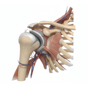
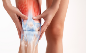
Pathologie – Was passiert bei Arthrose im Gelenk?
Bei Arthrose kommt es zu strukturellen Veränderungen im Gelenk, die jedoch nicht unbedingt mit Schmerzen verbunden sind. Die Schmerzursache liegt oft in Nervenstrukturen wie der Knochenhaut oder entzündetem Gewebe, nicht in den Knochen oder dem Gelenkknorpel selbst. Die Entstehung von Schmerzen bei Arthrose ist ein komplexer Prozess, der das zentrale und periphere Nervensystem involviert [3], [4], [5], [6].
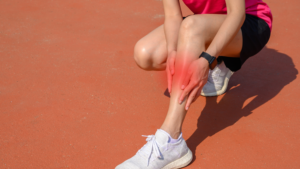
Krepitationen im Gelenk – Mythos 1: Gelenkgeräusche sind gefährlich
Krepitationen, also hörbare Gelenkgeräusche wie Knirschen oder Knacken, können oft Ängste auslösen. Jedoch zeigen Studien, dass diese Geräusche allein kein Indikator für den Schweregrad der Arthrose oder die Schmerzintensität sind [21], [22], [23], [24].
Wer ist davon betroffen? Mythos 2: Gelenkschmerzen sind altersbedingt
Während das Alter ein Risikofaktor für Arthrose ist, spielen auch andere Faktoren eine Rolle, darunter genetische Veranlagung, Verletzungen und Übergewicht. Es ist nicht korrekt anzunehmen, dass jeder automatisch im Alter unter Arthrose leidet [25], [26], [27].
Einteilung und Klassifikation – Mythos 3 & 4: Je größer der Gelenkschaden, desto schlimmer die Schmerzen; Arthrose tut wirklich immer weh
Das Ausmaß von Gelenkschäden korreliert nicht immer direkt mit der Schmerzintensität. Studien haben gezeigt, dass einige Personen radiologische Anzeichen von Arthrose aufweisen können, ohne dabei Symptome zu zeigen. Es besteht also nicht zwangsläufig ein direkter Zusammenhang zwischen dem Grad der Arthrose und dem Schmerzerleben [28], [29], [30].
Es ist von entscheidender Bedeutung, die gängigen Mythen rund um Arthrose zu entlarven und ein umfassendes Verständnis für diese Erkrankung zu fördern. In unserer Academy bieten wir vertiefende Einblicke und Lösungsansätze zur Bewältigung von Arthrose.
Prävention (Mythos 5: Arthrose lässt sich nur schwer vorbeugen)
Arthrose verursacht nicht nur persönliches Leid, sondern auch erhebliche Kosten im Gesundheitswesen. Um dies zu reduzieren, sind präventive Maßnahmen wichtig. Studien zeigen, dass sogar eine geringe Gewichtsabnahme das Risiko für Hüftgelenkersatz bei Hüftschmerzen reduzieren kann. Auch schmerzangepasstes Training ist entscheidend für Prävention und Behandlung [42].
Behandlung (Mythos 6 & 7: Sport ist Mord; Bei Arthrose sollte man Gelenke nur bewegen, nicht belasten; Von Arthrose betroffene Gelenke soll man schonen)
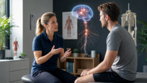
Aktive Therapieansätze sind essenziell, um ein aktives Leben trotz Arthrose zu ermöglichen. Multimodale Behandlungsstrategien, die Fitness, Stimmung, Stressabbau und Gewichtskontrolle umfassen, sind effektiver als einzelne Medikamente. Bewegungstherapie, insbesondere unter Anleitung, hat sich als wirksam erwiesen und verbessert Schmerzen und Funktion [1].
Konservative vs. operative Therapie (Mythos 8: Konservative Therapie hilft nur selten)
Konservative Behandlungen wie Hyaluronsäure-Injektionen können wirksam sein, insbesondere wenn andere Optionen nicht ausreichend sind. Operative Eingriffe wie eine Totalendoprothese sollten jedoch erst in Betracht gezogen werden, wenn alle anderen Optionen erschöpft sind und der Leidensdruck hoch ist [41], [45].
Fazit
Eine aktive Lebensweise und regelmäßiges, schmerzadaptiertes Training sind entscheidend, um mit Arthrose umzugehen und Lebensqualität zu erhalten. Individuelle Therapieansätze, Prävention, Bewegung und ganzheitliche Behandlungsmethoden, können dazu beitragen, die Auswirkungen von Arthrose zu minimieren und ein aktives Leben zu führen.
Dein DK Sports & Physio Team aus der Karlsruher Oststadt
Den ausführlichen Artikel findest du in unserer DK Academy.
Wir geben Physiotherapeuten, Trainern und allen Wissbegierigen einen sachlichen Einblick in die Physiotherapie und helfen so die Rehabilitation und das Training nach Verletzungen oder Beschwerden effizienter zu gestalten.
Sichere dir vollen Zugriff auf unsere Rehab Live Sessions, exklusive Review- und Blogartikel, Simple Tipps und Infografiken.

Du benötigst Physiotherapie im Raum Karlsruhe? Dann sind wir gerne für dich da und unterstützen dich!
Dein DK Sports & Physio Team aus der Karlsruher Oststadt
Unsere weiteren Blog-Artikel
Rotator cuff related shoulder pain – was steckt dahinter?
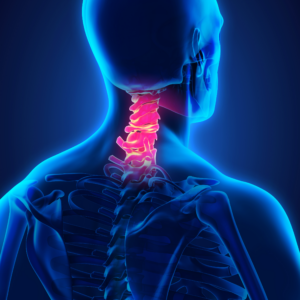
Hals über Kopf ins Märchen: Wer hat Angst vorm Handynacken?
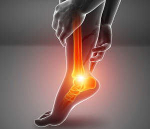
Plantarfasziitis oder Plantarfasziopathie – Ursachen und Lösungen bei Fersenschmerzen
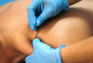
Vorsicht, piekst ein bisschen! Die Evidenz hinter Dry Needling
Was ist eine Arthrofibrose und was kann man dagegen tun?
Schmerzen erklären? Für Physios (k)ein großes Problem!
Osgood-Schlatter: Wenn Wachstumsschmerzen den Sport ausbremsen
Quellenangaben:
[1] S. L. Kolasinski u. a., „2019 American College of Rheumatology/Arthritis Foundation Guideline for the Management of Osteoarthritis of the Hand, Hip, and Knee“, Arthritis Rheumatol., Bd. 72, Nr. 2, S. 220–233, Feb. 2020, doi: 10.1002/art.41142.
[2] RKI, „12-Monats-Prävalenz von Arthrose in Deutschland“, 2017, doi: 10.17886/RKI-GBE-2017-054.
[3] R. Sen und J. A. Hurley, „Osteoarthritis“, in StatPearls, Treasure Island (FL): StatPearls Publishing, 2023. Zugegriffen: 26. Juni 2023. [Online]. Verfügbar unter: http://www.ncbi.nlm.nih.gov/books/NBK482326/
[4] L. Dr. Howard, „10 Recommendations To Thrive With Osteoarthritis of the Knee“. https://www.physio-network.com/blog/10-recommendations-to-thrive-with-osteoarthritis-of-the-knee/
[5] M. Imamura u. a., „Impact of nervous system hyperalgesia on pain, disability, and quality of life in patients with knee osteoarthritis: A controlled analysis“, Arthritis Rheum., Bd. 59, Nr. 10, S. 1424–1431, Okt. 2008, doi: 10.1002/art.24120.
[6] C. J. Woolf, „Pain: Moving from Symptom Control toward Mechanism-Specific Pharmacologic Management“, Ann. Intern. Med., Bd. 140, Nr. 6, S. 441, März 2004, doi: 10.7326/0003-4819-140-8-200404200-00010.
[7] R. Staud, „Are tender point injections beneficial: the role of tonic nociception in fibromyalgia“, Curr. Pharm. Des., Bd. 12, Nr. 1, S. 23–27, 2006.
[8] R. Staud und M. Spaeth, „Psychophysical and Neurochemical Abnormalities of Pain Processing in Fibromyalgia“, CNS Spectr., Bd. 13, Nr. S5, S. 12–17, März 2008, doi: 10.1017/S109285290002678X.
[9] R. Staud, R. C. Cannon, A. P. Mauderli, M. E. Robinson, D. D. Price, und C. J. Vierck, „Temporal summation of pain from mechanical stimulation of muscle tissue in normal controls and subjects with fibromyalgia syndrome“, Pain, Bd. 102, Nr. 1, S. 87–95, März 2003, doi: 10.1016/s0304-3959(02)00344-5.
[10] A. M. Abeles, M. H. Pillinger, B. M. Solitar, und M. Abeles, „Narrative Review: The Pathophysiology of Fibromyalgia“, Ann. Intern. Med., Bd. 146, Nr. 10, S. 726, Mai 2007, doi: 10.7326/0003-4819-146-10-200705150-00006.
[11] K. Lawson, „Treatment options and patient perspectives in the management of fibromyalgia: future trends“, Neuropsychiatr. Dis. Treat., S. 1059, Nov. 2008, doi: 10.2147/NDT.S3468.
[12] R. H. Gracely, F. Petzke, J. M. Wolf, und D. J. Clauw, „Functional magnetic resonance imaging evidence of augmented pain processing in fibromyalgia“, Arthritis Rheum., Bd. 46, Nr. 5, S. 1333–1343, Mai 2002, doi: 10.1002/art.10225.
[13] T. Giesecke u. a., „Evidence of augmented central pain processing in idiopathic chronic low back pain“, Arthritis Rheum., Bd. 50, Nr. 2, S. 613–623, Feb. 2004, doi: 10.1002/art.20063.
[14] S. E. Gwilym u. a., „Psychophysical and functional imaging evidence supporting the presence of central sensitization in a cohort of osteoarthritis patients“, Arthritis Rheum., Bd. 61, Nr. 9, S. 1226–1234, Sep. 2009, doi: 10.1002/art.24837.
[15] P. Creamer, M. Hunt, und P. Dieppe, „Pain mechanisms in osteoarthritis of the knee: effect of intraarticular anesthetic“, J. Rheumatol., Bd. 23, Nr. 6, S. 1031–1036, Juni 1996.
[16] M. Farrell, S. Gibson, J. McMeeken, und R. Helme, „Pain and hyperalgesia in osteoarthritis of the hands“, J. Rheumatol., Bd. 27, Nr. 2, S. 441–447, Feb. 2000.
[17] E. Kosek und G. Ordeberg, „Lack of pressure pain modulation by heterotopic noxious conditioning stimulation in patients with painful osteoarthritis before, but not following, surgical pain relief“, Pain, Bd. 88, Nr. 1, S. 69–78, Okt. 2000, doi: 10.1016/S0304-3959(00)00310-9.
[18] P. Bajaj, P. Bajaj, T. Graven-Nielsen, und L. Arendt-Nielsen, „Osteoarthritis and its association with muscle hyperalgesia: an experimental controlled study“, Pain, Bd. 93, Nr. 2, S. 107–114, Aug. 2001, doi: 10.1016/S0304-3959(01)00300-1.
[19] J. P. Shah, T. M. Phillips, J. V. Danoff, und L. H. Gerber, „An in vivo microanalytical technique for measuring the local biochemical milieu of human skeletal muscle“, J. Appl. Physiol., Bd. 99, Nr. 5, S. 1977–1984, Nov. 2005, doi: 10.1152/japplphysiol.00419.2005.
[20] J. P. Shah u. a., „Biochemicals Associated With Pain and Inflammation are Elevated in Sites Near to and Remote From Active Myofascial Trigger Points“, Arch. Phys. Med. Rehabil., Bd. 89, Nr. 1, S. 16–23, Jan. 2008, doi: 10.1016/j.apmr.2007.10.018.
[21] C. J. Robertson, M. Hurley, und F. Jones, „People’s beliefs about the meaning of crepitus in patellofemoral pain and the impact of these beliefs on their behaviour: A qualitative study“, Musculoskelet. Sci. Pract., Bd. 28, S. 59–64, Apr. 2017, doi: 10.1016/j.msksp.2017.01.012.
[22] M. F. Pazzinatto u. a., „What are the clinical implications of knee crepitus to individuals with knee osteoarthritis? An observational study with data from the Osteoarthritis Initiative“, Braz. J. Phys. Ther., Bd. 23, Nr. 6, S. 491–496, Nov. 2019, doi: 10.1016/j.bjpt.2018.11.001.
[23] J. Cibere u. a., „Association of clinical findings with pre-radiographic and radiographic knee osteoarthritis in a population-based study“, Arthritis Care Res., Bd. 62, Nr. 12, S. 1691–1698, Dez. 2010, doi: 10.1002/acr.20314.
[24] G. H. Lo, M. T. Strayhorn, J. B. Driban, L. L. Price, C. B. Eaton, und T. E. Mcalindon, „Subjective Crepitus as a Risk Factor for Incident Symptomatic Knee Osteoarthritis: Data From the Osteoarthritis Initiative“, Arthritis Care Res., Bd. 70, Nr. 1, S. 53–60, Jan. 2018, doi: 10.1002/acr.23246.
[25] B. Abramoff und F. E. Caldera, „Osteoarthritis“, Med. Clin. North Am., Bd. 104, Nr. 2, S. 293–311, März 2020, doi: 10.1016/j.mcna.2019.10.007.
[26] J. Ho, C. Mak, V. Sharma, K. To, und W. Khan, „Mendelian Randomization Studies of Lifestyle-Related Risk Factors for Osteoarthritis: A PRISMA Review and Meta-Analysis“, Int. J. Mol. Sci., Bd. 23, Nr. 19, S. 11906, Okt. 2022, doi: 10.3390/ijms231911906.
[27] F. Berenbaum, I. J. Wallace, D. E. Lieberman, und D. T. Felson, „Modern-day environmental factors in the pathogenesis of osteoarthritis“, Nat. Rev. Rheumatol., Bd. 14, Nr. 11, S. 674–681, Nov. 2018, doi: 10.1038/s41584-018-0073-x.
[28] J. H. Kellgren und J. S. Lawrence, „Radiological Assessment of Osteo-Arthrosis“, Ann. Rheum. Dis., Bd. 16, Nr. 4, S. 494–502, Dez. 1957, doi: 10.1136/ard.16.4.494.
[29] E. Halilaj, Y. Le, J. L. Hicks, T. J. Hastie, und S. L. Delp, „Modeling and predicting osteoarthritis progression: data from the osteoarthritis initiative“, Osteoarthritis Cartilage, Bd. 26, Nr. 12, S. 1643–1650, Dez. 2018, doi: 10.1016/j.joca.2018.08.003.
[30] M. T. Hannan, D. T. Felson, und T. Pincus, „Analysis of the discordance between radiographic changes and knee pain in osteoarthritis of the knee“, J. Rheumatol., Bd. 27, Nr. 6, S. 1513–1517, Juni 2000.
[31] A. G. Culvenor, B. E. Øiestad, H. F. Hart, J. J. Stefanik, A. Guermazi, und K. M. Crossley, „Prevalence of knee osteoarthritis features on magnetic resonance imaging in asymptomatic uninjured adults: a systematic review and meta-analysis“, Br. J. Sports Med., Bd. 53, Nr. 20, S. 1268–1278, Okt. 2019, doi: 10.1136/bjsports-2018-099257.
[32] Q. Weng u. a., „Comparative efficacy of exercise therapy and oral non-steroidal anti-inflammatory drugs and paracetamol for knee or hip osteoarthritis: a network meta-analysis of randomised controlled trials“, Br. J. Sports Med., S. bjsports-2022-105898, Jan. 2023, doi: 10.1136/bjsports-2022-105898.
[33] H. Bliddal, A. R. Leeds, und R. Christensen, „Osteoarthritis, obesity and weight loss: evidence, hypotheses and horizons – a scoping review“, Obes. Rev., Bd. 15, Nr. 7, S. 578–586, Juli 2014, doi: 10.1111/obr.12173.
[34] S. P. Messier u. a., „Intentional Weight Loss in Overweight and Obese Patients With Knee Osteoarthritis: Is More Better?“, Arthritis Care Res., Bd. 70, Nr. 11, S. 1569–1575, Nov. 2018, doi: 10.1002/acr.23608.
[35] M. M. Saw, T. Kruger-Jakins, N. Edries, und R. Parker, „Significant improvements in pain after a six-week physiotherapist-led exercise and education intervention, in patients with osteoarthritis awaiting arthroplasty, in South Africa: a randomised controlled trial“, BMC Musculoskelet. Disord., Bd. 17, Nr. 1, S. 236, Dez. 2016, doi: 10.1186/s12891-016-1088-6.
[36] R. Sasaki u. a., „Effect of exercise and/or educational interventions on physical activity and pain in patients with hip/knee osteoarthritis: A systematic review with meta-analysis“, PLOS ONE, Bd. 17, Nr. 11, S. e0275591, Nov. 2022, doi: 10.1371/journal.pone.0275591.
[37] I. Svege, L. Nordsletten, L. Fernandes, und M. A. Risberg, „Exercise therapy may postpone total hip replacement surgery in patients with hip osteoarthritis: a long-term follow-up of a randomised trial“, Ann. Rheum. Dis., Bd. 74, Nr. 1, S. 164–169, Jan. 2015, doi: 10.1136/annrheumdis-2013-203628.
[38] M. Schencking, S. Wilm, und M. Redaelli, „A comparison of Kneipp hydrotherapy with conventional physiotherapy in the treatment of osteoarthritis: a pilot trial“, J. Integr. Med., Bd. 11, Nr. 1, S. 17–25, Jan. 2013, doi: 10.3736/jintegrmed2013004.
[39] K. K. Sampath, R. Mani, T. Miyamori, und S. Tumilty, „The effects of manual therapy or exercise therapy or both in people with hip osteoarthritis: a systematic review and meta-analysis“, Clin. Rehabil., Bd. 30, Nr. 12, S. 1141–1155, Dez. 2016, doi: 10.1177/0269215515622670.
[40] N. Runge, A. Aina, und S. May, „The Benefits of Adding Manual Therapy to Exercise Therapy for Improving Pain and Function in Patients With Knee or Hip Osteoarthritis: A Systematic Review With Meta-analysis“, J. Orthop. Sports Phys. Ther., Bd. 52, Nr. 10, S. 675-A13, Okt. 2022, doi: 10.2519/jospt.2022.11062.
[41] T. V. Pereira u. a., „Viscosupplementation for knee osteoarthritis: systematic review and meta-analysis“, BMJ, S. e069722, Juli 2022, doi: 10.1136/bmj-2022-069722.
[42] Matziolis, G. M. & Deutsche Gesellschaft für Orthopädie und Orthopädische Chirurgie (DGOOC). (2019, 8. Juli). S2k-Leitlinie Koxarthrose. AWMF online Portal der wissenschaftlichen Medizin. Abgerufen am 5. Juli 2023, von https://register.awmf.org/de/leitlinien/detail/187-049 (Ursprünglich veröffentlicht 1997)
[43] Stöve, J. S. & Deutsche Gesellschaft für Orthopädie und Orthopädische Chirurgie (DGOOC). (2018, 18. Januar). S2k-Leitlinie Gonarthrose. AWMF online Das Portal der wissenschaftlichen Medizin. Abgerufen am 27. Juni 2023, von https://register.awmf.org/de/leitlinien/detail/187-050
[44] Osteoarthritis in over 16s: diagnosis and management. (2022, 19. Oktober). National Institute for Health and Care Excellence. Abgerufen am 5. Juli 2023, von https://www.nice.org.uk/guidance/ng226
[45] Deutschen Gesellschaft für Orthopädie und Unfallchirurgie e.V. (DGOU): Evidenz- und konsensbasierte Indikationskriterien zur Hüfttotalendoprothese bei Coxarthrose (EKIT-Hüfte). Version 1.0 (24.03.2021). Abgerufen am 5. August 2023, von https://www.awmf.org/leitlinien/detail/ll/187-001.html