

Einleitung: Wenn Therapie mehr braucht als Standardlösungen
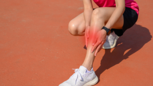
Schmerzen und Verletzungen gehören zum Alltag vieler Menschen und verschwinden häufig von selbst. Problematisch wird es jedoch bei langwierigen Beschwerden des Bewegungsapparates, die eine gezielte physiotherapeutische Behandlung erfordern. Genau hier entscheidet sich, wie erfolgreich deine Therapie wirklich ist – nicht nur durch Techniken, sondern durch ein ganzheitliches Verständnis von Patient:innen, Prozessen und Kommunikation.
Erfolgsfaktor 1: Die richtige Diagnose und Struktur
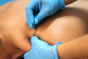
Eine erfolgreiche Physiotherapie beginnt mit einer fundierten Analyse. Es reicht nicht aus, sich ausschließlich auf ärztliche Diagnosen zu verlassen. Vielmehr ist es entscheidend, eigene Befunde zu erheben und die Beschwerden individuell zu verstehen.
Dabei geht es nicht nur um die körperliche Struktur, sondern auch um funktionelle Einschränkungen und Alltagsprobleme. Eine klare Struktur im Therapieprozess sorgt dafür, dass Maßnahmen gezielt eingesetzt werden und Fortschritte messbar bleiben.
Erfolgsfaktor 2: Individuelle Therapie statt Schema F
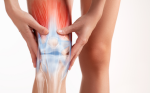
Jeder Patient bringt unterschiedliche Voraussetzungen, Ziele und Belastungen mit. Erfolgreiche Therapie bedeutet daher, standardisierte Behandlungen zu hinterfragen und individuell anzupassen.
Das betrifft sowohl die Auswahl der Übungen als auch deren Intensität und Progression. Eine gute Therapie orientiert sich immer daran, was für die jeweilige Person sinnvoll und realistisch umsetzbar ist – nicht daran, was theoretisch „ideal“ wäre.
Erfolgsfaktor 3: Aktive Einbindung der Patient:innen
Ein zentraler Bestandteil erfolgreicher Physiotherapie ist die aktive Mitarbeit der Patient:innen. Passive Maßnahmen allein führen selten zu nachhaltigem Erfolg.
Stattdessen sollten Patient:innen verstehen, warum sie bestimmte Übungen durchführen und welchen Einfluss ihr Verhalten auf den Heilungsverlauf hat. Ziel ist es, Eigenverantwortung zu fördern und langfristige Veränderungen im Alltag zu etablieren.
Erfolgsfaktor 4: Kommunikation als Schlüssel
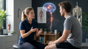
Die Art und Weise, wie du mit Patient:innen sprichst, hat einen erheblichen Einfluss auf den Therapieerfolg. Klare, verständliche und motivierende Kommunikation schafft Vertrauen und stärkt die Therapieadhärenz.
Dabei geht es nicht nur um Informationen, sondern auch um Erwartungen, Ängste und Überzeugungen. Wer es schafft, komplexe Inhalte einfach zu erklären und Sicherheit zu vermitteln, verbessert die Erfolgschancen der Behandlung deutlich.
Erfolgsfaktor 5: Evidenzbasierte Praxis
Moderne Physiotherapie basiert auf wissenschaftlichen Erkenntnissen. Es reicht nicht aus, sich auf Erfahrung oder traditionelle Methoden zu verlassen.
Therapeut:innen sollten regelmäßig neue Studien und Entwicklungen verfolgen und diese sinnvoll in ihre Praxis integrieren. Gleichzeitig gilt es, wissenschaftliche Erkenntnisse kritisch zu bewerten und individuell anzuwenden.
Erfolgsfaktor 6: Langfristige Perspektive statt kurzfristiger Lösungen
Viele Therapien scheitern daran, dass sie nur kurzfristige Verbesserungen anstreben. Nachhaltiger Erfolg entsteht jedoch erst, wenn Patient:innen langfristig belastbarer werden und eigenständig mit ihren Beschwerden umgehen können.
Das bedeutet, Therapie als Prozess zu verstehen, der über einzelne Sitzungen hinausgeht. Ziel ist nicht nur Schmerzreduktion, sondern eine dauerhafte Verbesserung von Funktion und Lebensqualität.
Fazit: Erfolgreiche Physiotherapie ist mehrdimensional
Eine erfolgreiche physiotherapeutische Behandlung entsteht aus dem Zusammenspiel mehrerer Faktoren: fundierte Diagnostik, individuelle Therapieplanung, aktive Patientenbeteiligung, effektive Kommunikation und wissenschaftliche Grundlage.
Wer diese Aspekte berücksichtigt, schafft nicht nur bessere Ergebnisse, sondern auch nachhaltige Veränderungen im Leben der Patient:innen.
Dein DK Sports & Physio Team aus der Karlsruher Oststadt
Den ausführlichen Artikel findest du in unserer DK Academy.
Wir geben Physiotherapeuten, Trainern und allen Wissbegierigen einen sachlichen Einblick in die Physiotherapie und helfen so die Rehabilitation und das Training nach Verletzungen oder Beschwerden effizienter zu gestalten.
Sichere dir vollen Zugriff auf unsere Rehab Live Sessions, exklusive Review- und Blogartikel, Simple Tipps und Infografiken.
Du benötigst Physiotherapie im Raum Karlsruhe? Dann sind wir gerne für dich da und unterstützen dich!
Dein DK Sports & Physio Team aus der Karlsruher Oststadt
Unsere weiteren Blog-Artikel
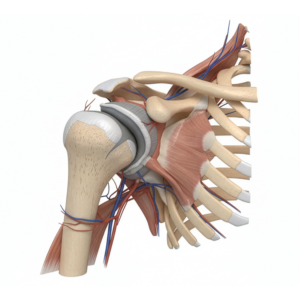
Rotator cuff related shoulder pain – was steckt dahinter?
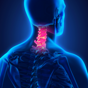
Hals über Kopf ins Märchen: Wer hat Angst vorm Handynacken?
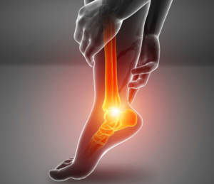
Plantarfasziitis oder Plantarfasziopathie – Ursachen und Lösungen bei Fersenschmerzen
Vorsicht, piekst ein bisschen! Die Evidenz hinter Dry Needling
Was ist eine Arthrofibrose und was kann man dagegen tun?
Schmerzen erklären? Für Physios (k)ein großes Problem!
Osgood-Schlatter: Wenn Wachstumsschmerzen den Sport ausbremsen
Quellenangaben:
[1] J. R. Andrews, G. L. Harrelson, und K. E. Wilk, Hrsg., Physical rehabilitation of the injured athlete, 4th edition. Philadelphia, PA: Elsevier/Saunders, 2012.
[2] V. K. Goradia, M. C. Rochat, W. A. Grana, M. D. Rohrer, und H. S. Prasad, „Tendon-to-bone healing of a semitendinosus tendon autograft used for ACL reconstruction in a sheep model“, Am. J. Knee Surg., Bd. 13, Nr. 3, S. 143–151, 2000.
[3] R. P. A. Janssen und S. U. Scheffler, „Intra-articular remodelling of hamstring tendon grafts after anterior cruciate ligament reconstruction“, Knee Surg. Sports Traumatol. Arthrosc., Bd. 22, Nr. 9, S. 2102–2108, Sep. 2014, doi: 10.1007/s00167-013-2634-5.
[4] S. Claes, P. Verdonk, R. Forsyth, und J. Bellemans, „The “Ligamentization” Process in Anterior Cruciate Ligament Reconstruction: What Happens to the Human Graft? A Systematic Review of the Literature“, Am. J. Sports Med., Bd. 39, Nr. 11, S. 2476–2483, Nov. 2011, doi: 10.1177/0363546511402662.
[5] S. U. Scheffler, F. N. Unterhauser, und A. Weiler, „Graft remodeling and ligamentization after cruciate ligament reconstruction“, Knee Surg. Sports Traumatol. Arthrosc., Bd. 16, Nr. 9, S. 834–842, Sep. 2008, doi: 10.1007/s00167-008-0560-8.
[6] L. Moretti, D. Bizzoca, G. D. Cassano, N. Caringella, M. Delmedico, und B. Moretti, „Graft Intra-Articular Remodeling and Bone Incorporation in ACL Reconstruction: The State of the Art and Clinical Implications“, J. Clin. Med., Bd. 11, Nr. 22, S. 6704, Nov. 2022, doi: 10.3390/jcm11226704.
[7] C. J. Hadley u. a., „Safer Return to Play After Anterior Cruciate Ligament Reconstruction: Evaluation of a Return-to-Play Checklist“, Orthop. J. Sports Med., Bd. 10, Nr. 4, S. 232596712210904, Apr. 2022, doi: 10.1177/23259671221090412.
[8] G. Radovanović, S. Bohm, K. K. Peper, A. Arampatzis, und K. Legerlotz, „Evidence-Based High-Loading Tendon Exercise for 12 Weeks Leads to Increased Tendon Stiffness and Cross-Sectional Area in Achilles Tendinopathy: A Controlled Clinical Trial“, Sports Med. – Open, Bd. 8, Nr. 1, S. 149, Dez. 2022, doi: 10.1186/s40798-022-00545-5.
[9] H. Alfredson, T. Pietilä, P. Jonsson, und R. Lorentzon, „Heavy-Load Eccentric Calf Muscle Training For the Treatment of Chronic Achilles Tendinosis“, Am. J. Sports Med., Bd. 26, Nr. 3, S. 360–366, Mai 1998, doi: 10.1177/03635465980260030301.
[10] T. B. Cardoso, T. Pizzari, R. Kinsella, D. Hope, und J. L. Cook, „Current trends in tendinopathy management“, Best Pract. Res. Clin. Rheumatol., Bd. 33, Nr. 1, S. 122–140, Feb. 2019, doi: 10.1016/j.berh.2019.02.001.
[11] T. Saueressig u. a., „Primary surgery versus primary rehabilitation for treating anterior cruciate ligament injuries: a living systematic review and meta-analysis“, Br. J. Sports Med., S. bjsports-2021-105359, Aug. 2022, doi: 10.1136/bjsports-2021-105359.
[12] J. Hayden, M. W. Van Tulder, A. Malmivaara, und B. W. Koes, „Exercise therapy for treatment of non-specific low back pain“, Cochrane Database Syst. Rev., Bd. 2011, Nr. 2, Juli 2005, doi: 10.1002/14651858.CD000335.pub2.
[13] E. Vermeire, H. Hearnshaw, P. Van Royen, und J. Denekens, „Patient adherence to treatment: three decades of research. A comprehensive review“, J. Clin. Pharm. Ther., Bd. 26, Nr. 5, S. 331–342, Okt. 2001, doi: 10.1046/j.1365-2710.2001.00363.x.
[14] L. M. Vasey, „DNAs and DNCTs — Why Do Patients Fail to Begin or to Complete a Course of Physiotherapy Treatment?“, Physiotherapy, Bd. 76, Nr. 9, S. 575–578, Sep. 1990, doi: 10.1016/S0031-9406(10)63052-0.
[15] M. Friedrich, G. Gittler, Y. Halberstadt, T. Cermak, und I. Heiller, „Combined exercise and motivation program: Effect on the compliance and level of disability of patients with chronic low back pain: A randomized controlled trial“, Arch. Phys. Med. Rehabil., Bd. 79, Nr. 5, S. 475–487, Mai 1998, doi: 10.1016/S0003-9993(98)90059-4.
[16] R. Campbell, „Why don’t patients do their exercises? Understanding non-compliance with physiotherapy in patients with osteoarthritis of the knee“, J. Epidemiol. Community Health, Bd. 55, Nr. 2, S. 132–138, Feb. 2001, doi: 10.1136/jech.55.2.132.
[17] E. M. Sluijs, G. J. Kok, und J. Van Der Zee, „Correlates of Exercise Compliance in Physical Therapy“, Phys. Ther., Bd. 73, Nr. 11, S. 771–782, Nov. 1993, doi: 10.1093/ptj/73.11.771.
[18] K. Jack, S. M. McLean, J. K. Moffett, und E. Gardiner, „Barriers to treatment adherence in physiotherapy outpatient clinics: A systematic review“, Man. Ther., Bd. 15, Nr. 3, S. 220–228, Juni 2010, doi: 10.1016/j.math.2009.12.004.
[19] R. S. Miller, „Inattentive and contented: Relationship commitment and attention to alternatives.“, J. Pers. Soc. Psychol., Bd. 73, Nr. 4, S. 758–766, Okt. 1997, doi: 10.1037/0022-3514.73.4.758.
[20] R. Chester, C. Jerosch-Herold, J. Lewis, und L. Shepstone, „Psychological factors are associated with the outcome of physiotherapy for people with shoulder pain: a multicentre longitudinal cohort study“, Br. J. Sports Med., Bd. 52, Nr. 4, S. 269–275, Feb. 2018, doi: 10.1136/bjsports-2016-096084.
[21] D. Kalauokalani, D. C. Cherkin, K. J. Sherman, T. D. Koepsell, und R. A. Deyo, „Lessons from a Trial of Acupuncture and Massage for Low Back Pain: Patient Expectations and Treatment Effects“, Spine, Bd. 26, Nr. 13, S. 1418–1424, Juli 2001, doi: 10.1097/00007632-200107010-00005.
[22] N. E. Foster, „Beliefs and preferences: do they help determine the outcome of musculoskeletal problems?“, Phys. Ther. Rev., Bd. 12, Nr. 3, S. 199–206, Sep. 2007, doi: 10.1179/108331907X222976.
[23] Preference Collaborative Review Group, „Patients’ preferences within randomised trials: systematic review and patient level meta-analysis“, BMJ, Bd. 337, Nr. oct31 1, S. a1864–a1864, Okt. 2008, doi: 10.1136/bmj.a1864.
[24] M. V. Mondloch, D. C. Cole, und J. W. Frank, „Does how you do depend on how you think you’ll do? A systematic review of the evidence for a relation between patients’ recovery expectations and health outcomes“, CMAJ Can. Med. Assoc. J. J. Assoc. Medicale Can., Bd. 165, Nr. 2, S. 174–179, Juli 2001.
[25] K. J. Sherman u. a., „Treatment expectations and preferences as predictors of outcome of acupuncture for chronic back pain“, Spine, Bd. 35, Nr. 15, S. 1471–1477, Juli 2010, doi: 10.1097/BRS.0b013e3181c2a8d3.
[26] M. Müller-Schrader u. a., „Individual treatment expectations predict clinical outcome after lumbar injections against low back pain“, Pain, Bd. 164, Nr. 1, S. 132–141, Jan. 2023, doi: 10.1097/j.pain.0000000000002674.
[27] K. Schmidt u. a., „The beneficial effect of positive treatment expectations on pharmacological migraine prophylaxis“, Pain, Bd. 163, Nr. 2, S. e319–e327, Feb. 2022, doi: 10.1097/j.pain.0000000000002341.
[28] T. D. Wager u. a., „Placebo-Induced Changes in fMRI in the Anticipation and Experience of Pain“, Science, Bd. 303, Nr. 5661, S. 1162–1167, Feb. 2004, doi: 10.1126/science.1093065.
[29] L. Y. Atlas und T. D. Wager, „How expectations shape pain“, Neurosci. Lett., Bd. 520, Nr. 2, S. 140–148, Juni 2012, doi: 10.1016/j.neulet.2012.03.039.
[30] C. A. Porro u. a., „Does Anticipation of Pain Affect Cortical Nociceptive Systems?“, J. Neurosci., Bd. 22, Nr. 8, S. 3206–3214, Apr. 2002, doi: 10.1523/JNEUROSCI.22-08-03206.2002.
[31] J. Lorenz u. a., „Cortical correlates of false expectations during pain intensity judgments—a possible manifestation of placebo/nocebo cognitions☆“, Brain. Behav. Immun., Bd. 19, Nr. 4, S. 283–295, Juli 2005, doi: 10.1016/j.bbi.2005.03.010.
[32] A. Ploghaus, L. Becerra, C. Borras, und D. Borsook, „Neural circuitry underlying pain modulation: expectation, hypnosis, placebo“, Trends Cogn. Sci., Bd. 7, Nr. 5, S. 197–200, Mai 2003, doi: 10.1016/S1364-6613(03)00061-5.
[33] M. Corbetta und G. L. Shulman, „Control of goal-directed and stimulus-driven attention in the brain“, Nat. Rev. Neurosci., Bd. 3, Nr. 3, S. 201–215, März 2002, doi: 10.1038/nrn755.
[34] J. Weiss u. a., „Loss-of-function mutations in sodium channel Nav1.7 cause anosmia“, Nature, Bd. 472, Nr. 7342, S. 186–190, Apr. 2011, doi: 10.1038/nature09975.
[35] J. J. Cox u. a., „An SCN9A channelopathy causes congenital inability to experience pain“, Nature, Bd. 444, Nr. 7121, S. 894–898, Dez. 2006, doi: 10.1038/nature05413.
[36] M. Buckthorpe, A. Tamisari, und F. D. Villa, „A TEN TASK-BASED PROGRESSION IN REHABILITATION AFTER ACL RECONSTRUCTION: FROM POST-SURGERY TO RETURN TO PLAY – A CLINICAL COMMENTARY“, Int. J. Sports Phys. Ther., Bd. 15, Nr. 4, S. 611–623, Mai 2020, doi: 10.26603/ijspt20200611.
[37] A. Serner u. a., „Return to Sport After Criteria-Based Rehabilitation of Acute Adductor Injuries in Male Athletes: A Prospective Cohort Study“, Orthop. J. Sports Med., Bd. 8, Nr. 1, S. 232596711989724, Jan. 2020, doi: 10.1177/2325967119897247.
[38] M. Artus, D. A. van der Windt, K. P. Jordan, und E. M. Hay, „Low back pain symptoms show a similar pattern of improvement following a wide range of primary care treatments: a systematic review of randomized clinical trials“, Rheumatology, Bd. 49, Nr. 12, S. 2346–2356, Dez. 2010, doi: 10.1093/rheumatology/keq245.
[39] R. Leproult, „Effect of 1 Week of Sleep Restriction on Testosterone Levels in Young Healthy Men“, JAMA, Bd. 305, Nr. 21, S. 2173, Juni 2011, doi: 10.1001/jama.2011.710.
[40] W. J. Bremner, „Testosterone Deficiency and Replacement in Older Men“, N. Engl. J. Med., Bd. 363, Nr. 2, S. 189–191, Juli 2010, doi: 10.1056/NEJMe1006197.
[41] S. Cohen, W. J. Doyle, C. M. Alper, D. Janicki-Deverts, und R. B. Turner, „Sleep Habits and Susceptibility to the Common Cold“, Arch. Intern. Med., Bd. 169, Nr. 1, S. 62, Jan. 2009, doi: 10.1001/archinternmed.2008.505.
[42] M. D. Milewski u. a., „Chronic Lack of Sleep is Associated With Increased Sports Injuries in Adolescent Athletes“, J. Pediatr. Orthop., Bd. 34, Nr. 2, S. 129–133, März 2014, doi: 10.1097/BPO.0000000000000151.
[43] M. Schrimpf, G. Liegl, M. Boeckle, A. Leitner, P. Geisler, und C. Pieh, „The effect of sleep deprivation on pain perception in healthy subjects: a meta-analysis“, Sleep Med., Bd. 16, Nr. 11, S. 1313–1320, Nov. 2015, doi: 10.1016/j.sleep.2015.07.022.
[44] S. Schuh-Hofer u. a., „One night of total sleep deprivation promotes a state of generalized hyperalgesia: A surrogate pain model to study the relationship of insomnia and pain“, Pain, Bd. 154, Nr. 9, S. 1613–1621, Sep. 2013, doi: 10.1016/j.pain.2013.04.046.
[45] Y. H. Chiu u. a., „Poor sleep and depression are independently associated with a reduced pain threshold. Results of a population based study“, Pain, Bd. 115, Nr. 3, S. 316–321, Juni 2005, doi: 10.1016/j.pain.2005.03.009.
[46] M. Y. Aǧargün, I. Tekeoǧlu, A. Güneş, B. Adak, H. Kara, und M. Ercan, „Sleep quality and pain threshold in patients with fibromyalgia“, Compr. Psychiatry, Bd. 40, Nr. 3, S. 226–228, Mai 1999, doi: 10.1016/S0010-440X(99)90008-1.
[47] P. Meerlo, A. Sgoifo, und D. Suchecki, „Restricted and disrupted sleep: Effects on autonomic function, neuroendocrine stress systems and stress responsivity“, Sleep Med. Rev., Bd. 12, Nr. 3, S. 197–210, Juni 2008, doi: 10.1016/j.smrv.2007.07.007.
[48] L. Morselli, R. Leproult, M. Balbo, und K. Spiegel, „Role of sleep duration in the regulation of glucose metabolism and appetite“, Best Pract. Res. Clin. Endocrinol. Metab., Bd. 24, Nr. 5, S. 687–702, Okt. 2010, doi: 10.1016/j.beem.2010.07.005.
[49] K. L. Knutson, K. Spiegel, P. Penev, und E. Van Cauter, „The metabolic consequences of sleep deprivation“, Sleep Med. Rev., Bd. 11, Nr. 3, S. 163–178, Juni 2007, doi: 10.1016/j.smrv.2007.01.002.
[50] F. Atrooz und S. Salim, „Sleep deprivation, oxidative stress and inflammation“, in Advances in Protein Chemistry and Structural Biology, Bd. 119, Elsevier, 2020, S. 309–336. doi: 10.1016/bs.apcsb.2019.03.001.
[51] H. H. K. Fullagar und J. D. Bartlett, „Time to wake up: individualising the approach to sleep promotion interventions“, Br. J. Sports Med., Bd. 50, Nr. 3, S. 143–144, Feb. 2016, doi: 10.1136/bjsports-2015-095759.
[52] K. De Pauw, B. Roelands, U. Marušič, H. F. Tellez, K. Knaepen, und R. Meeusen, „Brain mapping after prolonged cycling and during recovery in the heat“, J. Appl. Physiol., Bd. 115, Nr. 9, S. 1324–1331, Nov. 2013, doi: 10.1152/japplphysiol.00633.2013.
[53] R. Naylor, C. Hayes, und G. Egger, „The Relationship Between Lifestyle, Metaflammation, and Chronic Pain: A Systematic Review“, Am. J. Lifestyle Med., Bd. 7, Nr. 2, S. 130–137, März 2013, doi: 10.1177/1559827612451710.
[54] A. Okifuji und B. Hare, „The association between chronic pain and obesity“, J. Pain Res., S. 399, Juli 2015, doi: 10.2147/JPR.S55598.
[55] K. D. Tipton, E. Borsheim, S. E. Wolf, A. P. Sanford, und R. R. Wolfe, „Acute response of net muscle protein balance reflects 24-h balance after exercise and amino acid ingestion“, Am. J. Physiol.-Endocrinol. Metab., Bd. 284, Nr. 1, S. E76–E89, Jan. 2003, doi: 10.1152/ajpendo.00234.2002.
[56] K. A. Reich, Y.-W. Chen, P. D. Thompson, E. P. Hoffman, und P. M. Clarkson, „Forty-eight hours of unloading and 24 h of reloading lead to changes in global gene expression patterns related to ubiquitination and oxidative stress in humans“, J. Appl. Physiol., Bd. 109, Nr. 5, S. 1404–1415, Nov. 2010, doi: 10.1152/japplphysiol.00444.2010.
[57] K. D. Tipton, „Nutritional Support for Exercise-Induced Injuries“, Sports Med. Auckl. NZ, Bd. 45 Suppl 1, S. S93-104, Nov. 2015, doi: 10.1007/s40279-015-0398-4.
[58] A. A. Aragon u. a., „International society of sports nutrition position stand: diets and body composition“, J. Int. Soc. Sports Nutr., Bd. 14, Nr. 1, S. 16, Dez. 2017, doi: 10.1186/s12970-017-0174-y.
[59] D. K. Layman, „Protein Quantity and Quality at Levels above the RDA Improves Adult Weight Loss“, J. Am. Coll. Nutr., Bd. 23, Nr. sup6, S. 631S-636S, Dez. 2004, doi: 10.1080/07315724.2004.10719435.
[60] D. K. Layman, „Dietary Guidelines should reflect new understandings about adult protein needs“, Nutr. Metab., Bd. 6, Nr. 1, S. 12, 2009, doi: 10.1186/1743-7075-6-12.
[61] D. S. Weigle u. a., „A high-protein diet induces sustained reductions in appetite, ad libitum caloric intake, and body weight despite compensatory changes in diurnal plasma leptin and ghrelin concentrations“, Am. J. Clin. Nutr., Bd. 82, Nr. 1, S. 41–48, Juli 2005, doi: 10.1093/ajcn/82.1.41.
[62] E. B. Parr u. a., „Alcohol ingestion impairs maximal post-exercise rates of myofibrillar protein synthesis following a single bout of concurrent training“, PloS One, Bd. 9, Nr. 2, S. e88384, 2014, doi: 10.1371/journal.pone.0088384.
[63] M. K. Jung u. a., „Alcohol exposure and mechanisms of tissue injury and repair“, Alcohol. Clin. Exp. Res., Bd. 35, Nr. 3, S. 392–399, März 2011, doi: 10.1111/j.1530-0277.2010.01356.x.
[64] A. G. Shaw, S. Chae, D. E. Levitt, J. L. Nicholson, J. L. Vingren, und D. W. Hill, „Effect of Previous-Day Alcohol Ingestion on Muscle Function and Performance of Severe-Intensity Exercise“, Int. J. Sports Physiol. Perform., Bd. 17, Nr. 1, S. 44–49, Jan. 2022, doi: 10.1123/ijspp.2020-0790.
[65] D. Zheng u. a., „Alcohol consumption and sleep quality: a community-based study“, Public Health Nutr., Bd. 24, Nr. 15, S. 4851–4858, Okt. 2021, doi: 10.1017/S1368980020004553.
[66] I. M. Colrain, C. L. Nicholas, und F. C. Baker, „Alcohol and the sleeping brain“, in Handbook of Clinical Neurology, Bd. 125, Elsevier, 2014, S. 415–431. doi: 10.1016/B978-0-444-62619-6.00024-0.
[67] D. C. Evans, R. G. Martindale, L. N. Kiraly, und C. M. Jones, „Nutrition optimization prior to surgery“, Nutr. Clin. Pract. Off. Publ. Am. Soc. Parenter. Enter. Nutr., Bd. 29, Nr. 1, S. 10–21, Feb. 2014, doi: 10.1177/0884533613517006.
[68] R. H. Demling, „Nutrition, anabolism, and the wound healing process: an overview“, Eplasty, Bd. 9, S. e9, 2009.
[69] R. Jäger u. a., „International Society of Sports Nutrition Position Stand: protein and exercise“, J. Int. Soc. Sports Nutr., Bd. 14, S. 20, 2017, doi: 10.1186/s12970-017-0177-8.
[70] D. W. Hart u. a., „Efficacy of a high-carbohydrate diet in catabolic illness“, Crit. Care Med., Bd. 29, Nr. 7, S. 1318–1324, Juli 2001, doi: 10.1097/00003246-200107000-00004.
[71] D. T. Thomas, K. A. Erdman, und L. M. Burke, „American College of Sports Medicine Joint Position Statement. Nutrition and Athletic Performance“, Med. Sci. Sports Exerc., Bd. 48, Nr. 3, S. 543–568, März 2016, doi: 10.1249/MSS.0000000000000852.
[72] J. K. Stechmiller, „Understanding the Role of Nutrition and Wound Healing“, Nutr. Clin. Pract., Bd. 25, Nr. 1, S. 61–68, Feb. 2010, doi: 10.1177/0884533609358997.
[73] E. Lin, J. G. Kotani, und S. F. Lowry, „Nutritional modulation of immunity and the inflammatory response“, Nutrition, Bd. 14, Nr. 6, S. 545–550, Juni 1998, doi: 10.1016/S0899-9007(98)00046-X.
[74] P. C. Calder, „n -3 Fatty acids, inflammation and immunity: new mechanisms to explain old actions“, Proc. Nutr. Soc., Bd. 72, Nr. 3, S. 326–336, Aug. 2013, doi: 10.1017/S0029665113001031.
[75] L. Galland, „Diet and Inflammation“, Nutr. Clin. Pract., Bd. 25, Nr. 6, S. 634–640, Dez. 2010, doi: 10.1177/0884533610385703.
[76] P. Agostoni u. a., „Metabolic exercise test data combined with cardiac and kidney indexes, the MECKI score: A multiparametric approach to heart failure prognosis“, Int. J. Cardiol., Bd. 167, Nr. 6, S. 2710–2718, Sep. 2013, doi: 10.1016/j.ijcard.2012.06.113.
[77] J. K. Kiecolt-Glaser, G. G. Page, P. T. Marucha, R. C. MacCallum, und R. Glaser, „Psychological influences on surgical recovery: Perspectives from psychoneuroimmunology.“, Am. Psychol., Bd. 53, Nr. 11, S. 1209–1218, 1998, doi: 10.1037/0003-066X.53.11.1209.
[78] P. H. Rosenberger, P. Jokl, und J. Ickovics, „Psychosocial Factors and Surgical Outcomes: An Evidence-Based Literature Review“:, J. Am. Acad. Orthop. Surg., Bd. 14, Nr. 7, S. 397–405, Juli 2006, doi: 10.5435/00124635-200607000-00002.
[79] A. Cole-King und K. G. Harding, „Psychological Factors and Delayed Healing in Chronic Wounds“:, Psychosom. Med., Bd. 63, Nr. 2, S. 216–220, März 2001, doi: 10.1097/00006842-200103000-00004.
[80] M. Ebrecht, J. Hextall, L.-G. Kirtley, A. Taylor, M. Dyson, und J. Weinman, „Perceived stress and cortisol levels predict speed of wound healing in healthy male adults“, Psychoneuroendocrinology, Bd. 29, Nr. 6, S. 798–809, Juli 2004, doi: 10.1016/S0306-4530(03)00144-6.
[81] L. McGuire u. a., „Pain and wound healing in surgical patients“, Ann. Behav. Med., Bd. 31, Nr. 2, S. 165–172, Apr. 2006, doi: 10.1207/s15324796abm3102_8.
[82] J. E. Graham, T. F. Robles, J. K. Kiecolt-Glaser, W. B. Malarkey, M. G. Bissell, und R. Glaser, „Hostility and pain are related to inflammation in older adults“, Brain. Behav. Immun., Bd. 20, Nr. 4, S. 389–400, Juli 2006, doi: 10.1016/j.bbi.2005.11.002.
[83] P. T. Marucha, J. K. Kiecolt-Glaser, und M. Favagehi, „Mucosal Wound Healing Is Impaired by Examination Stress“:, Psychosom. Med., Bd. 60, Nr. 3, S. 362–365, 1998, doi: 10.1097/00006842-199805000-00025.
[84] J. A. Bosch, C. G. Engeland, J. T. Cacioppo, und P. T. Marucha, „Depressive Symptoms Predict Mucosal Wound Healing“, Psychosom. Med., Bd. 69, Nr. 7, S. 597–605, Sep. 2007, doi: 10.1097/PSY.0b013e318148c682.
[85] B. Johnstone und M. T. Bayliss, „The Large Proteoglycans of the Human Intervertebral Disc: Changes in Their Biosynthesis and Structure with Age, Topography, and Pathology“, Spine, Bd. 20, Nr. 6, S. 674–684, März 1995, doi: 10.1097/00007632-199503150-00008.
[86] G. H. Montgomery, D. David, G. Winkel, J. H. Silverstein, und D. H. Bovbjerg, „The Effectiveness of Adjunctive Hypnosis with Surgical Patients: A Meta-Analysis“:, Anesth. Analg., Bd. 94, Nr. 6, S. 1639–1645, Juni 2002, doi: 10.1097/00000539-200206000-00052.
[87] J. A. Blumenthal u. a., „Long-term Effects of Exercise on Psychological Functioning in Older Men and Women“, J. Gerontol., Bd. 46, Nr. 6, S. P352–P361, Nov. 1991, doi: 10.1093/geronj/46.6.P352.
[88] C. F. Emery, J. K. Kiecolt-Glaser, R. Glaser, W. B. Malarkey, und D. J. Frid, „Exercise Accelerates Wound Healing Among Healthy Older Adults: A Preliminary Investigation“, J. Gerontol. A. Biol. Sci. Med. Sci., Bd. 60, Nr. 11, S. 1432–1436, Nov. 2005, doi: 10.1093/gerona/60.11.1432.
[89] D. A. Padgett und R. Glaser, „How stress influences the immune response“, Trends Immunol., Bd. 24, Nr. 8, S. 444–448, Aug. 2003, doi: 10.1016/S1471-4906(03)00173-X.
[90] R. Glaser und J. K. Kiecolt-Glaser, „Stress-induced immune dysfunction: implications for health“, Nat. Rev. Immunol., Bd. 5, Nr. 3, S. 243–251, März 2005, doi: 10.1038/nri1571.
[91] J.-P. Gouin und J. K. Kiecolt-Glaser, „The Impact of Psychological Stress on Wound Healing: Methods and Mechanisms“, Immunol. Allergy Clin. North Am., Bd. 31, Nr. 1, S. 81–93, Feb. 2011, doi: 10.1016/j.iac.2010.09.010.
[92] A. Steptoe, J. Wardle, T. M. Pollard, L. Canaan, und G. J. Davies, „Stress, social support and health-related behavior: A study of smoking, alcohol consumption and physical exercise“, J. Psychosom. Res., Bd. 41, Nr. 2, S. 171–180, Aug. 1996, doi: 10.1016/0022-3999(96)00095-5.
[93] P. P. Vitaliano, J. M. Scanlan, J. Zhang, M. V. Savage, I. B. Hirsch, und I. C. Siegler, „A Path Model of Chronic Stress, the Metabolic Syndrome, and Coronary Heart Disease“:, Psychosom. Med., Bd. 64, Nr. 3, S. 418–435, Mai 2002, doi: 10.1097/00006842-200205000-00006.
[94] K. Benveniste und P. Thut, „The Effect of Chronic Alcoholism on Wound Healing“, Exp. Biol. Med., Bd. 166, Nr. 4, S. 568–575, Apr. 1981, doi: 10.3181/00379727-166-41110.
[95] P. Silverstein, „Smoking and wound healing“, Am. J. Med., Bd. 93, Nr. 1, S. S22–S24, Juli 1992, doi: 10.1016/0002-9343(92)90623-J.
[96] M. Altemus, B. Rao, F. S. Dhabhar, W. Ding, und R. D. Granstein, „Stress-Induced Changes in Skin Barrier Function in Healthy Women“, J. Invest. Dermatol., Bd. 117, Nr. 2, S. 309–317, Aug. 2001, doi: 10.1046/j.1523-1747.2001.01373.x.
[97] J. D. Veldhuis und A. Iranmanesh, „Physiological Regulation of the Human Growth Hormone (GH)-Insulin-Like Growth Factor Type I (IGF-I) Axis: Predominant Impact of Age, Obesity, Gonadal Function, and Sleep“, Sleep, Bd. 19, Nr. suppl_10, S. S221–S224, Dez. 1996, doi: 10.1093/sleep/19.suppl_10.S221.
[98] K. T. Keylock, V. J. Vieira, M. A. Wallig, L. A. DiPietro, M. Schrementi, und J. A. Woods, „Exercise accelerates cutaneous wound healing and decreases wound inflammation in aged mice“, Am. J. Physiol.-Regul. Integr. Comp. Physiol., Bd. 294, Nr. 1, S. R179–R184, Jan. 2008, doi: 10.1152/ajpregu.00177.2007.
[99] L. Russell, „The importance of patients’ nutritional status in wound healing“, Br. J. Nurs., Bd. 10, Nr. Sup1, S. S42–S49, März 2001, doi: 10.12968/bjon.2001.10.Sup1.5336.
[100] M. E. Posthauer, „The Role of Nutrition in Wound Care“:, Adv. Skin Wound Care, Bd. 19, Nr. 1, S. 43–52, Jan. 2006, doi: 10.1097/00129334-200601000-00015.
[101] J. C. McDaniel, M. Belury, K. Ahijevych, und W. Blakely, „Omega‐3 fatty acids effect on wound healing“, Wound Repair Regen., Bd. 16, Nr. 3, S. 337–345, Mai 2008, doi: 10.1111/j.1524-475X.2008.00388.x.
[102] D. L. Sackett, W. M. C. Rosenberg, J. A. M. Gray, R. B. Haynes, und W. S. Richardson, „Evidence based medicine: what it is and what it isn’t“, BMJ, Bd. 312, Nr. 7023, S. 71–72, Jan. 1996, doi: 10.1136/bmj.312.7023.71.