Wie schief bin ich eigentlich und ist das schlimm?

Muskuloskelettale Beschwerden, besonders in der Beckenregion, sind ein häufiges Thema in der medizinischen Praxis. Häufig wird dabei auf strukturelle und posturale Probleme wie ein schiefes Becken, Beinlängendifferenzen oder das Iliosakralgelenk (ISG) verwiesen, um Schmerzen zu erklären. Das sogenannte posturale, strukturelle und biomechanische Modell (PSB) dominiert seit Jahrzehnten die Diagnostik und Therapieansätze in diesem Bereich. Doch ist dieses Modell wirklich tragfähig?
Das PSB-Paradigma auf dem Prüfstand
Das PSB-Modell stützt sich stark auf palpatorische Untersuchungen und manuelle Tests, um potenzielle Asymmetrien und Pathologien zu identifizieren. Die Reliabilität (Zuverlässigkeit) und Validität (Gültigkeit) solcher Tests ist jedoch fragwürdig. Studien zeigen, dass die Fähigkeit, anatomische Strukturen zuverlässig zu ertasten, oft unzureichend ist. Dies stellt die klinische Relevanz solcher Diagnosen infrage.
Palpation und Beinlängendifferenzen
Die Palpation spielt eine zentrale Rolle im PSB-Modell, jedoch weisen zahlreiche Studien auf die geringe Verlässlichkeit dieser Methode hin. So konnte in Untersuchungen keine ausreichende Reproduzierbarkeit von Tastbefunden festgestellt werden, was bedeutet, dass viele der palpatorischen Diagnosen ungenau sind. Auch Beinlängendifferenzen werden oft als Ursache für Beschwerden genannt, doch Studien zeigen, dass fast jeder Mensch unterschiedlich lange Beine hat, ohne dabei Schmerzen zu empfinden. Zudem sind die Messmethoden für Beinlängendifferenzen meist unzuverlässig und liefern keine konsistenten Ergebnisse.
Der Mythos Iliosakralgelenk (ISG)
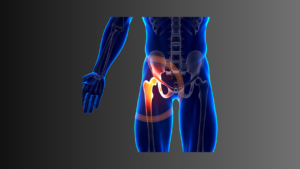
Das ISG steht häufig im Fokus von Diagnosen bei Beckenschmerzen. Tests zur Beweglichkeit und Schmerzprovokation des ISG weisen jedoch ebenfalls methodische Mängel auf. Studien zeigen, dass das ISG kaum Bewegungen zulässt und selbst bei Hyperbeweglichkeit kein Zusammenhang zu Schmerzen besteht. Die diagnostischen Tests für das ISG sind daher kaum valide und sollten kritisch hinterfragt werden.
Das untere gekreuzte Syndrom (Pelvic Tilt)
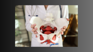
Das „untere gekreuzte Syndrom“ beschreibt eine Kippung des Beckens, die oft mit muskulären Dysbalancen und Schmerzen in Verbindung gebracht wird. Doch auch hier zeigt die wissenschaftliche Literatur, dass muskuläre Ungleichgewichte nicht verlässlich identifiziert werden können. Studien belegen zudem, dass die Beckenstellung kaum eine Rolle bei der Entstehung von Rückenschmerzen spielt, da viele Menschen eine Beckenkippung aufweisen, ohne Beschwerden zu haben.
Zusammenfassung
Die weit verbreiteten Annahmen des PSB-Modells sollten kritisch hinterfragt werden. Die Reliabilität und Validität vieler manueller Tests und palpatorischer Befunde sind unzureichend. Anstatt Ressourcen in Diagnosen zu investieren, deren Relevanz wissenschaftlich nicht belegt ist, sollten sich Therapeutinnen mehr auf funktionelle Aspekte und die bio-psycho-soziale Natur von Schmerzen konzentrieren. Eine Neuausrichtung hin zu evidenzbasierten Ansätzen könnte die Behandlungsstrategien und die Versorgung von Patientinnen verbessern.
Dein DK Sports & Physio Team aus Karlsruhe
Den ausführlichen Artikel findest du in unserer DK Academy.
Wir geben Physiotherapeuten, Trainern und allen Wissbegierigen einen sachlichen Einblick in die Physiotherapie und helfen so die Rehabilitation und das Training nach Verletzungen oder Beschwerden effizienter zu gestalten.
Sichere dir vollen Zugriff auf unsere Rehab Live Sessions, exklusive Review- und Blogartikel, Simple Tipps und Infografiken.

Du benötigst Physiotherapie im Raum Karlsruhe? Dann sind wir gerne für dich da und unterstützen dich!
Dein DK Sports & Physio Team aus der Karlsruher Oststadt
Unsere weiteren Blog-Artikel


Was manuelle Lymphdrainage (nicht) kann

Gelenkgeräusche – muss man sich Gedanken machen?

Seitlicher Hüftschmerz Update und Deep-Dive

Faszinierend oder fast nicht belegt – Der Faszien-Hype kritisch betrachtet

Von Eisbädern bis Foam Rolling: Was hilft wirklich gegen Muskelkater?

Kreuzbandrehabilitation – Wie lange brauche ich die Unterarmgehstützen?

Wie schief bin ich eigentlich und ist das schlimm?
Quellenangaben:
[1] J. Setchell, N. Costa, M. Ferreira, J. Makovey, M. Nielsen, and P. W. Hodges, “Individuals’ explanations for their persistent or recurrent low back pain: a cross-sectional survey,” BMC Musculoskelet. Disord., vol. 18, no. 1, p. 466, Nov. 2017, doi: 10.1186/s12891-017-1831-7.
[2] E. Lederman, “The fall of the postural-structural-biomechanical model in manual and physical therapies: exemplified by lower back pain,” J. Bodyw. Mov. Ther., vol. 15, no. 2, pp. 131–138, Apr. 2011, doi: 10.1016/j.jbmt.2011.01.011.
[3] S. J. Kamper, “Reliability and Validity: Linking Evidence to Practice,” J. Orthop. Sports Phys. Ther., vol. 49, no. 4, pp. 286–287, Apr. 2019, doi: 10.2519/jospt.2019.0702.
[4] B. A. Stovall and S. Kumar, “Reliability of Bony Anatomic Landmark Asymmetry Assessment in the Lumbopelvic Region: Application to Osteopathic Medical Education,” J. Am. Osteopath. Assoc., vol. 110, no. 11, pp. 667–674, Nov. 2010.
[5] T. Paulet and G. Fryer, “Inter-examiner reliability of palpation for tissue texture abnormality in the thoracic paraspinal region,” Int. J. Osteopath. Med., vol. 12, no. 3, pp. 92–96, Sep. 2009, doi: 10.1016/j.ijosm.2008.07.001.
[6] P. S. Nolet et al., “Reliability and validity of manual palpation for the assessment of patients with low back pain: a systematic and critical review,” Chiropr. Man. Ther., vol. 29, no. 1, p. 33, Aug. 2021, doi: 10.1186/s12998-021-00384-3.
[7] T. Liem, “Pitfalls and challenges involved in the process of perception and interpretation of palpatory findings,” Int. J. Osteopath. Med., vol. 17, no. 4, pp. 243–249, Dec. 2014, doi: 10.1016/j.ijosm.2014.04.005.
[8] M. Alfuth, P. Fichter, and A. Knicker, “Leg length discrepancy: A systematic review on the validity and reliability of clinical assessments and imaging diagnostics used in clinical practice,” PLoS ONE, vol. 16, no. 12, p. e0261457, Dec. 2021, doi: 10.1371/journal.pone.0261457.
[9] E. Gomez-Aguilar, M. Reina-Bueno, G. Lafuente-Sotillos, R. Montes-Salas, P. V. Munuera-Martinez, and J. M. Castillo-Lopez, “Validity of clinical methods in the detection of leg-length discrepancies,” Hip Int. J. Clin. Exp. Res. Hip Pathol. Ther., vol. 31, no. 2, pp. 186–190, Mar. 2021, doi: 10.1177/1120700020910108.
[10] G. A. Knutson, “Anatomic and functional leg-length inequality: A review and recommendation for clinical decision-making. Part I, anatomic leg-length inequality: prevalence, magnitude, effects and clinical significance,” Chiropr. Osteopat., vol. 13, p. 11, Jul. 2005, doi: 10.1186/1746-1340-13-11.
[11] S. O’Brien, G. Kernohan, C. Fitzpatrick, J. Hill, and D. Beverland, “Perception of imposed leg length inequality in normal subjects,” Hip Int. J. Clin. Exp. Res. Hip Pathol. Ther., vol. 20, no. 4, pp. 505–511, 2010, doi: 10.1177/112070001002000414.
[12] P. van der Wurff, R. H. Hagmeijer, and W. Meyne, “Clinical tests of the sacroiliac joint. A systematic methodological review. Part 1: Reliability,” Man. Ther., vol. 5, no. 1, pp. 30–36, Feb. 2000, doi: 10.1054/math.1999.0228.
[13] P. van der Wurff, W. Meyne, and R. H. Hagmeijer, “Clinical tests of the sacroiliac joint,” Man. Ther., vol. 5, no. 2, pp. 89–96, May 2000, doi: 10.1054/math.1999.0229.
[14] D. L. Riddle and J. K. Freburger, “Evaluation of the presence of sacroiliac joint region dysfunction using a combination of tests: a multicenter intertester reliability study,” Phys. Ther., vol. 82, no. 8, pp. 772–781, Aug. 2002.
[15] U. Holmgren and K. Waling, “Inter-examiner reliability of four static palpation tests used for assessing pelvic dysfunction,” Man. Ther., vol. 13, no. 1, pp. 50–56, Feb. 2008, doi: 10.1016/j.math.2006.09.009.
[16] B. Sturesson, A. Uden, and A. Vleeming, “A radiostereometric analysis of the movements of the sacroiliac joints in the reciprocal straddle position,” Spine, vol. 25, no. 2, pp. 214–217, Jan. 2000, doi: 10.1097/00007632-200001150-00012.
[17] B. Sturesson, A. Uden, and A. Vleeming, “A radiostereometric analysis of movements of the sacroiliac joints during the standing hip flexion test,” Spine, vol. 25, no. 3, pp. 364–368, Feb. 2000, doi: 10.1097/00007632-200002010-00018.
[18] T. Tullberg, S. Blomberg, B. Branth, and R. Johnsson, “Manipulation does not alter the position of the sacroiliac joint. A roentgen stereophotogrammetric analysis,” Spine, vol. 23, no. 10, pp. 1124–1128; discussion 1129, May 1998, doi: 10.1097/00007632-199805150-00010.
[19] D. de F. A. de Toledo, F. B. Kochem, and J. G. Silva, “High-velocity, low-amplitude manipulation (HVLA) does not alter three-dimensional position of sacroiliac joint in healthy men: A quasi-experimental study,” J. Bodyw. Mov. Ther., vol. 24, no. 1, pp. 190–193, Jan. 2020, doi: 10.1016/j.jbmt.2019.05.020.
[20] A. Goode, E. J. Hegedus, P. Sizer, J.-M. Brismee, A. Linberg, and C. E. Cook, “Three-Dimensional Movements of the Sacroiliac Joint: A Systematic Review of the Literature and Assessment of Clinical Utility,” J. Man. Manip. Ther., vol. 16, no. 1, pp. 25–38, 2008.
[21] T. J. Kibsgård, S. M. Röhrl, O. Røise, B. Sturesson, and B. Stuge, “Movement of the sacroiliac joint during the Active Straight Leg Raise test in patients with long-lasting severe sacroiliac joint pain,” Clin. Biomech. Bristol Avon, vol. 47, pp. 40–45, Aug. 2017, doi: 10.1016/j.clinbiomech.2017.05.014.
[22] T. J. Kibsgård, O. Røise, B. Sturesson, S. M. Röhrl, and B. Stuge, “Radiosteriometric analysis of movement in the sacroiliac joint during a single-leg stance in patients with long-lasting pelvic girdle pain,” Clin. Biomech. Bristol Avon, vol. 29, no. 4, pp. 406–411, Apr. 2014, doi: 10.1016/j.clinbiomech.2014.02.002.
[23] N. K. Vøllestad, P. A. Torjesen, and H. S. Robinson, “Association between the serum levels of relaxin and responses to the active straight leg raise test in pregnancy,” Man. Ther., vol. 17, no. 3, pp. 225–230, Jun. 2012, doi: 10.1016/j.math.2012.01.003.
[24] A. Vleeming, R. Stoeckart, A. C. Volkers, and C. J. Snijders, “Relation between form and function in the sacroiliac joint. Part I: Clinical anatomical aspects,” Spine, vol. 15, no. 2, pp. 130–132, Feb. 1990, doi: 10.1097/00007632-199002000-00016.
[25] C. J. Snijders, A. Vleeming, and R. Stoeckart, “Transfer of lumbosacral load to iliac bones and legs Part 1: Biomechanics of self-bracing of the sacroiliac joints and its significance for treatment and exercise,” Clin. Biomech. Bristol Avon, vol. 8, no. 6, pp. 285–294, Nov. 1993, doi: 10.1016/0268-0033(93)90002-Y.
[26] S. J. Slater and D. A. Barron, “Pelvic fractures-A guide to classification and management,” Eur. J. Radiol., vol. 74, no. 1, pp. 16–23, Apr. 2010, doi: 10.1016/j.ejrad.2010.01.025.
[27] S. J. Preece, P. Willan, C. J. Nester, P. Graham-Smith, L. Herrington, and P. Bowker, “Variation in pelvic morphology may prevent the identification of anterior pelvic tilt,” J. Man. Manip. Ther., vol. 16, no. 2, pp. 113–117, 2008, doi: 10.1179/106698108790818459.
[28] E. Frese, M. Brown, and B. J. Norton, “Clinical reliability of manual muscle testing. Middle trapezius and gluteus medius muscles,” Phys. Ther., vol. 67, no. 7, pp. 1072–1076, Jul. 1987, doi: 10.1093/ptj/67.7.1072.
[29] R. W. Bohannon, “Manual muscle testing: does it meet the standards of an adequate screening test?,” Clin. Rehabil., vol. 19, no. 6, pp. 662–667, Sep. 2005, doi: 10.1191/0269215505cr873oa.
[30] S. C. Cuthbert and G. J. Goodheart, “On the reliability and validity of manual muscle testing: a literature review,” Chiropr. Osteopat., vol. 15, p. 4, Mar. 2007, doi: 10.1186/1746-1340-15-4.
[31] M. L. Walker, J. M. Rothstein, S. D. Finucane, and R. L. Lamb, “Relationships between lumbar lordosis, pelvic tilt, and abdominal muscle performance,” Phys. Ther., vol. 67, no. 4, pp. 512–516, Apr. 1987, doi: 10.1093/ptj/67.4.512.
[32] Y. Li, P. W. McClure, and N. Pratt, “The effect of hamstring muscle stretching on standing posture and on lumbar and hip motions during forward bending,” Phys. Ther., vol. 76, no. 8, pp. 836–845; discussion 845-849, Aug. 1996, doi: 10.1093/ptj/76.8.836.
[33] B. J. Elliott, N. Hookway, B. M. Tate, and M. G. Hines, “Does passive hip stiffness or range of motion correlate with spinal curvature and posture during quiet standing?,” Gait Posture, vol. 85, pp. 273–279, Mar. 2021, doi: 10.1016/j.gaitpost.2021.02.012.
[34] L. Herrington, “Assessment of the degree of pelvic tilt within a normal asymptomatic population,” Man. Ther., vol. 16, no. 6, pp. 646–648, Dec. 2011, doi: 10.1016/j.math.2011.04.006.